. 2019 May;39(5):808-821.
doi: 10.1177/0271678X17733640. Epub 2017 Oct 19.

Recurrent moderate hypoglycemia exacerbates oxidative damage and neuronal death leading to cognitive dysfunction after the hypoglycemic coma

Affiliations

Recurrent moderate hypoglycemia exacerbates oxidative damage and neuronal death leading to cognitive dysfunction after the hypoglycemic coma

Gabriela Languren et al. J Cereb Blood Flow Metab. 2019 May.

Abstract

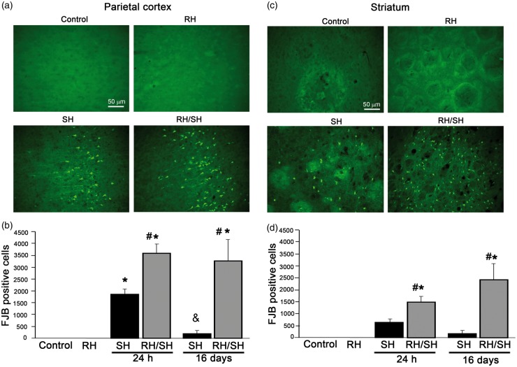
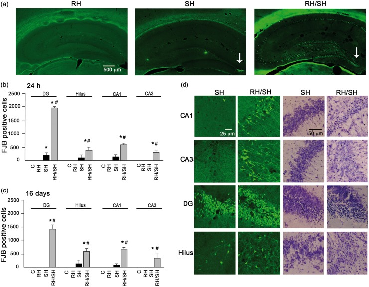
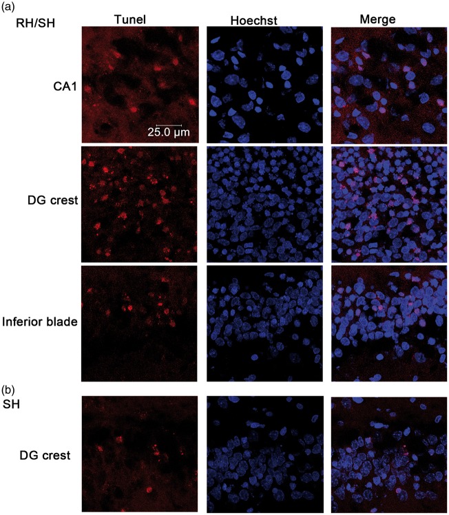
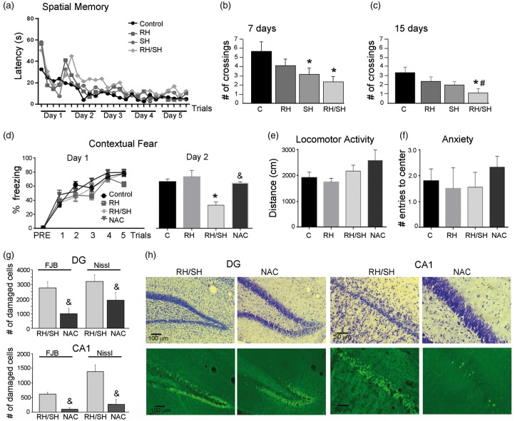
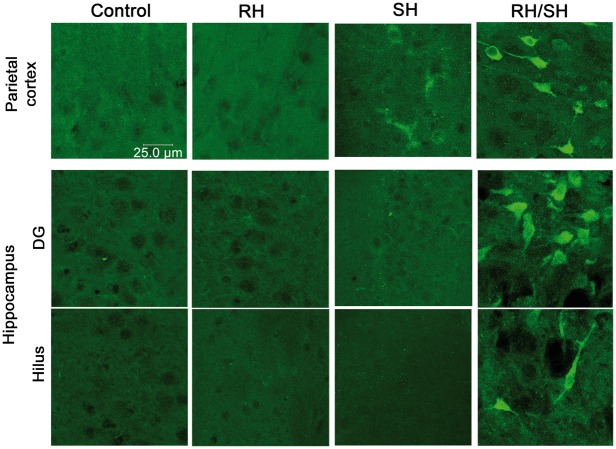
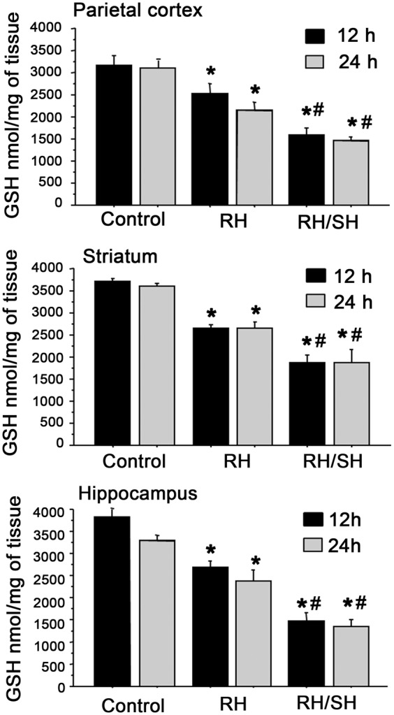
Moderate recurrent hypoglycemia (RH) is frequent in Type 1 diabetes mellitus (TIDM) patients who are under intensive insulin therapy increasing the risk for severe hypoglycemia (SH). The consequences of RH are not well understood and its repercussions on neuronal damage and cognitive function after a subsequent episode of SH have been poorly investigated. In the current study, we have addressed this question and observed that previous RH during seven consecutive days exacerbated oxidative damage and neuronal death induced by a subsequent episode of SH accompanied by a short period of coma, in the parietal cortex, the striatum and mainly in the hippocampus. These changes correlated with a severe decrease in reduced glutathione content (GSH), and a significant spatial and contextual memory deficit. Administration of the antioxidant, N-acetyl-L-cysteine, (NAC) reduced neuronal death and prevented cognitive impairment. These results demonstrate that previous RH enhances brain vulnerability to acute hypoglycemia and suggests that this effect is mediated by the decline in the antioxidant defense and oxidative damage. The present results highlight the importance of an adequate control of moderate hypoglycemic episodes in TIDM.

Keywords: Diabetes; antioxidants; cognitive impairment/decline; glucose; hippocampus; hypoglycemia; risk factors; selective neuronal death.


Figures

Figure 1.
Figure 1.
Neuronal death induced by the hypoglycemic coma in the parietal cortex and the striatum is exacerbated by antecedent RH. Neuronal death was evaluated 24 h and 16 days after the treatments and the number of FJB-positive cells was counted. Representative micrographs showing no FJB-positive in control and RH-treated animals and degenerating cells in the parietal cortex (a) and the striatum (c) of animals exposed to SH and RH/SH. (b–d) Cell death was exacerbated in animals exposed to RH/SH at 24 h and 16 days after the treatments. Data are expressed as mean ± SEM from seven animals per group. *p < 0.0001 relative to control and RH, #p < 0.0001 relative to SH.
Figure 2.
Figure 2.
Neuronal death induced by the hypoglycemic coma in the hippocampus is exacerbated by antecedent RH. Neuronal death was evaluated 24 h or 16 days after the different treatments in brain sections stained with Nissl or FJB. (a) Representative micrographs showing degenerating cells only in the crest of the DG of animals exposed to SH and extensive neuronal death in animals from the RH/SH group. (b–c) Quantitative data showing exacerbated neuronal death in animals exposed to RH/SH at 24 h or 16 days. (d) Representative micrographs showing FJB-positive and pyknotic cells in the different hippocampal subregions of rats exposed to the different treatments. Data are expressed as mean ± SEM from seven animals per group. *p < 0.0001 relative to control and RH, #p < 0.0001 relative to SH.
Figure 3.
Figure 3.
DNA fragmentation induced by the hypoglycemic coma is exacerbated by RH. The presence of DNA fragmentation was examined by the TUNEL assay and Hoechst was used as counterstain. (a) Representative micrographs showing TUNEL-positive cells in CA1 and the DG of rats treated with RH/SH. (b) In rats exposed to SH alone TUNEL-positive were detected only in the crest of the DG.
Figure 4.
Figure 4.
Previous RH aggravates cognitive impairment induced by the hypoglycemic coma. Spatial memory test (Water maze) was performed 7 and 15 days after the treatments. (a) Learning curve of animals exposed to the different treatments. (b) At day 7, animals of the SH and RH/SH groups showed a decrease in the number of crossings. (c) At 15 days, only animals of the RH/SH group showed a significant decrease in the number of crossings. Data are expressed as mean ± SEM from seven animals per group. *p < 0.01 relative to control #p < 0.05 relative to SH. Contextual fear conditioning test was performed 48 h after the treatments. (d) No statistical differences between the groups were observed in the acquisition phase (day 1). The RH/SH group showed a significant decrease, while the NAC group showed no change in the percent freezing relative to control and RH groups during retrieval (day 2). Data are expressed as mean ± SEM from six to nine animals per group. *p < 0.0001 relative to control and RH &p < 0.05 relative RH/SH. (e) No significant changes were observed between groups in (e) locomotor activity and (f) anxiety tests. (g) Number of damaged cells (FJB-positive group and pyknotic Nissl stained) in the dentate gyrus (DG) and CA1 of animals of the RS/SH and the NAC groups, 24 h after the fear-conditioning test. Data are expressed as mean ± SEM from four to six animals per group. &p < 0.002 relative to RH/SH). (h) Representative images showing the protective effect induced by NAC administration against cell damage (FJB-positive cell and pyknotic cells in Nissl stained section) induced in the DG and CA1 after exposure to RH/SH.
Figure 5.
Figure 5.
Immunoreactivity to 4-HNE in the brain of animals exposed to RH/SH. Representative fluorescent images showing the presence of 4-HNE-positive cells in (a) parietal cortex, (b) striatum and (c) hippocampus of animals treated with RH/SH. No 4-HNE-positive cells were detected in control, RH and SH groups.
Figure 6.
Figure 6.
Immunoreactivity to 3-NT in the brain of animals exposed to RH/SH. Representative fluorescent images showing the presence of 3-NT-positive cells in the parietal cortex and the hippocampus of animals treated with RH/SH. No 3-NT-positive cells were detected in control and RH groups and only a few cells were observed in the parietal cortex of animals of the SH groups.
Figure 7.
Figure 7.
Changes in GSH content after RH and RH/SH. Animals were exposed to the different treatments and 12 and 24 h later reduced GSH levels were measured in different brain regions. Animals exposed to RH show a significant moderate decrease in reduced GSH in all brain regions, which is further decreased in animals from the RH/SH group (n = 3–7 animals per group). Data are expressed in means ± SEM. *p < 0.01 relative to control; #p < 0.01 relative to RH.

References

    1. Donelly LA, Morris AD, Frier BM, et al. Frequency and predictors of hypoglycaemia in Type 1 and insulin-treated type 2 diabetes: a population-based study. Diab Med 2005; 22: 749–755. - PubMed
    1. Heller S, Colagiuri S, Vaaler S, et al. Hypoglycaemia with insulin aspart: a doubleblind randomised, crossover trial in subjects with Type 1 diabetes. Diab Med 2004; 21: 769–775. - PubMed
    1. Blasetti A, Di Giulio C, Tocco AM, et al. Variables associated with severe hypoglycemia in children and adolescents with Type 1 diabetes: a population-based study. Pediatr Diab 2011; 12: 4–10. - PubMed
    1. Frier BM. Hypoglycaemia in diabetes mellitus: epidemiology and clinical implications. Nat Rev Endocrinol 2014; 10: 711–722. - PubMed
    1. Languren G, Montiel T, Julio-Amilpas A, et al. Neuronal damage and cognitive impairment associated with hypoglycemia: an integrated view. Neurochem Int 2013; 63: 331–343. - PubMed

Publication types

LinkOut - more resources