Review
. 2017 Oct 18;24(1):80.
doi: 10.1186/s12929-017-0384-2.

The recent development and applications of fluidic channels by 3D printing

Affiliations
Review

The recent development and applications of fluidic channels by 3D printing

Yufeng Zhou. J Biomed Sci. .

Abstract

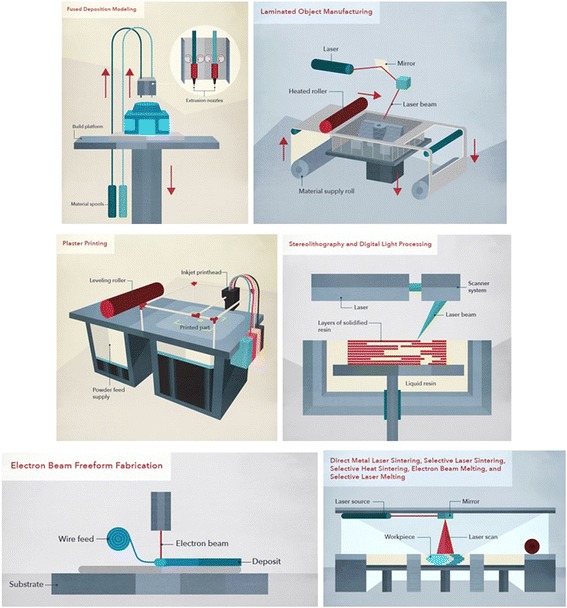
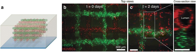
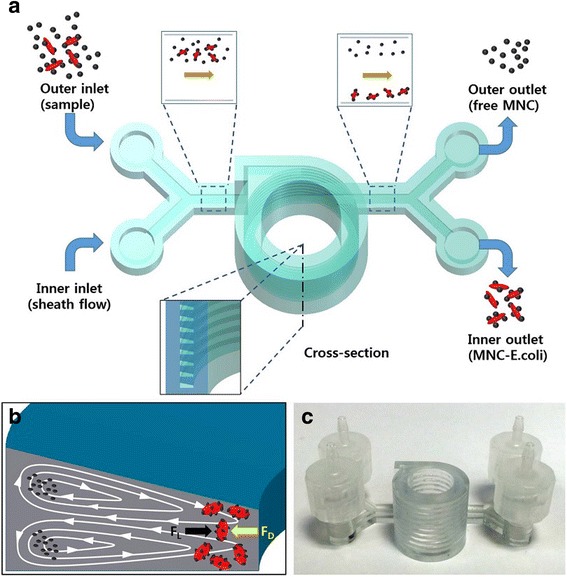
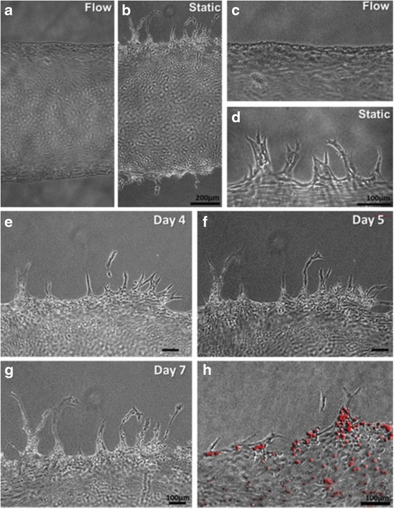
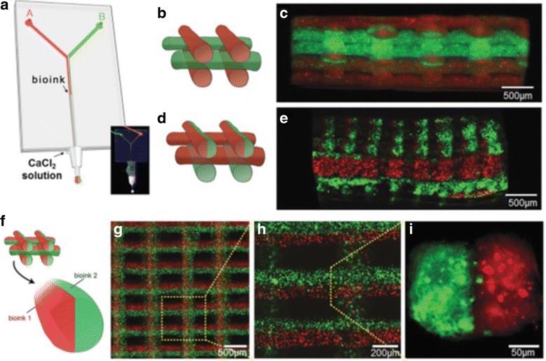
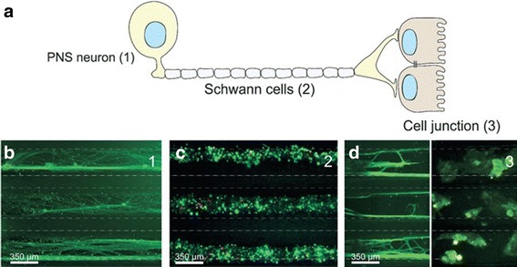
The technology of "Lab-on-a-Chip" allows the synthesis and analysis of chemicals and biological substance within a portable or handheld device. The 3D printed structures enable precise control of various geometries. The combination of these two technologies in recent years makes a significant progress. The current approaches of 3D printing, such as stereolithography, polyjet, and fused deposition modeling, are introduced. Their manufacture specifications, such as surface roughness, resolution, replication fidelity, cost, and fabrication time, are compared with each other. Finally, novel application of 3D printed channel in biology are reviewed, including pathogenic bacteria detection using magnetic nanoparticle clusters in a helical microchannel, cell stimulation by 3D chemical gradients, perfused functional vascular channels, 3D tissue construct, organ-on-a-chip, and miniaturized fluidic "reactionware" devices for chemical syntheses. Overall, the 3D printed fluidic chip is becoming a powerful tool in the both medical and chemical industries.

Keywords: 3D printing; Diagnosis; Fluidic channel; Lab-on-a-chip; Reactionware; Tissue engineering.


Conflict of interest statement

Ethics approval and consent to participate

Not applicable.

Consent for publication

Not applicable.

Competing interests

The author declares no competing interest.

Publisher’s Note

Springer Nature remains neutral with regard to jurisdictional claims in published maps and institutional affiliations.

Figures

Fig. 1
Fig. 1
Diagram of various 3D printing techniques including fusion deposition modelling, laminated object manufacturing, plaster printing, sterolithography, electrom beam freeform fabrication, and selective laser sintering, with courtesy of CustomMade.com
Fig. 2
Fig. 2
a Effect of the peeling direction and crossover features on the successful rate, scale bar of 250 μm, b image of the permanently healed 3D chip (basket-weaving configuration) loaded with yellow and blue dyes and magnified image of the dash line depicted region, with courtesy of [124]
Fig. 3
Fig. 3
a Fabrication procedures, 1: direct 2D or 3D sugar structures printing; 2: pouring PDMS on the structures; 3: removing sugar structures to obtain fluidic chips without further sealing, b printed 3D sugar structure, and c a 12-layer 3D microvascular network, with courtesy of [125]
Fig. 4
Fig. 4
Schematic diagrams and images of fabricating 3D microchannel using liquid metal: a printing the liquid metal, b encapsulating the printed pattern in a polymer, c withdrawing the metal, and (d) refilling with red dye solution, with courtesy of [126]
Fig. 5
Fig. 5
Schematic diagram of omnidirectional printing of 3D microvascular networks in a hydrogel reservoir: a deposition of a fugitive ink into a gel reservoir to pattern hierarchical, branching networks, b filling the voids induced by nozzle translation with liquid that migrates from the fluid capping layer, c yield of a chemically cross-linked, hydrogel matrix by photo-polymerizing the reservoir, d, e exposure of the microvascular channels by removing the liquefied ink under a modest vacuum, and f fluorescent image of a 3D microvascular network (scale bar = 10 mm), with courtesy of [53]
Fig. 6
Fig. 6
a Schematic view of red and green filaments with RFP HUVECs and GFP HNDF-laden GelMA ink, respectively, and (b, c) fluorescence images of an engineered tissue construct cultured for 0 and 2 days, respectively, with courtesy of [121]
Fig. 7
Fig. 7
a Illustration of separating bacteria by inertial focusing, b Dean vortices in a channel with trapezoid cross-section, and c photograph of the 3D printed device, with courtesy of [70]
Fig. 8
Fig. 8
Morphology of HUVECs on the vascular channel edge in a, c dynamic culture and b, d static culture on Day 5, e-g the sprouts budded from the channel wall extending during culture and maintaining filopodia-like protrusion on the tip in the static condition, and h Luminal structure of sprouts as confirmed by the injection of fluorescence microbeads (10 mm), with courtesy of [85]
Fig. 9
Fig. 9
a A microfluidic system with the bioinks flow containing red and green fluorescent beads, photograph (inset) of the coaxial needle system with a “Y”-shaped microchannel, the illustration and fluorescence image of 3D construct with b,c alternate, d,e alternate/simultaneous, and fi simultaneous deposition, with courtesy of [88]
Fig. 10
Fig. 10
a Schematic of a peripheral 3D neural systems-on-a-chip consisting of peripheral nervous system (PNS) neuron, Schwann cells, and cell junction, micrograph showing three parallel microchannels of b PNS neurons stained by green tau, c peripheral nerve fibres stained using tri-colour pseudorabies virus d axon termini (green tau stained) and epithelial cells stained by gree tau and cytokeratin, respectively, with courtesy of [127]

References

    1. Barry R, Ivanov D. Microfluidics in biotechnology. J Nanobiotechnol. 2004;2(1):2. - PMC - PubMed
    1. Dittrich PS, Manz A. Lab-on-a-chip: microfluidics in drug discovery. Nat Rev Drug Discov. 2006;5(3):210–218. - PubMed
    1. Guo L, Feng J, Fang Z, Xu J, Lu X. Application of microfluidic “lab-on-a-chip” for the detection of mycotoxins in foods. Trends Food Sci Technol. 2015;46(2):252–263.
    1. Weibel DB, Whitesides GM. Applications of microfluidics in chemical biology. Curr Opin Chem Biol. 2006;10(6):584–591. - PubMed
    1. Whitesides GM. The origins and the future of microfluidics. Nature. 2006;442(7101):368–373. - PubMed