Review
. 2017 Oct 19;18(10):2183.
doi: 10.3390/ijms18102183.

The Structure of Human Neuromuscular Junctions: Some Unanswered Molecular Questions

Affiliations
Review

The Structure of Human Neuromuscular Junctions: Some Unanswered Molecular Questions

Clarke R Slater. Int J Mol Sci. .

Abstract

The commands that control animal movement are transmitted from motor neurons to their target muscle cells at the neuromuscular junctions (NMJs). The NMJs contain many protein species whose role in transmission depends not only on their inherent properties, but also on how they are distributed within the complex structure of the motor nerve terminal and the postsynaptic muscle membrane. These molecules mediate evoked chemical transmitter release from the nerve and the action of that transmitter on the muscle. Human NMJs are among the smallest known and release the smallest number of transmitter "quanta". By contrast, they have the most deeply infolded postsynaptic membranes, which help to amplify transmitter action. The same structural features that distinguish human NMJs make them particularly susceptible to pathological processes. While much has been learned about the molecules which mediate transmitter release and action, little is known about the molecular processes that control the growth of the cellular and subcellular components of the NMJ so as to give rise to its mature form. A major challenge for molecular biologists is to understand the molecular basis for the development and maintenance of functionally important aspects of NMJ structure, and thereby to point to new directions for treatment of diseases in which neuromuscular transmission is impaired.

Keywords: disease; human; mouse; neuromuscular junction; neuromuscular transmission; structure.


Conflict of interest statement

The author declares no conflicts of interest.

Figures

Figure 1
Figure 1
Mammalian neuromuscular junctions (NMJ) structure. (A) Diagram showing a motor axon which branches to innervate two muscle fibers at the NMJs. Note the extensive branching of the motor axon terminal. Arrow shows region where nerve has been removed, revealing postsynaptic folds (From Salpeter, 1986, with permission) [2]; (B) Human NMJ showing the nerve terminal (green, immunolabeling of synaptophysin and neurofilament protein) and the postsynaptic acetylcholine receptors (AChRs) (red, fluorescent tagged α-bungarotoxin). Note the swollen “boutons” of the nerve terminal, from which transmitter is released. Scale bar, 20 μm; (C) Electron micrograph of a section through a single bouton. Note the extensive infolding of the postsynaptic muscle fiber membrane. Scale bar, 1 μm.
Figure 2
Figure 2
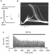
Basics of neuromuscular transmission. (A) Intracellular recordings of synaptic events at a mouse NMJ: top, miniature endplate potential (mEPP), the response to a single quantum of ACh; bottom, plate potential (EPP), the nerve-evoked response to about 50 quanta; (B) Intracellular recordings from an NMJ from a human patient with myasthenia gravis. The smaller events are subthreshold EPPs, which result from the low numbers of AChR in this disease. (From Elmqvist et al., 1964, with permission) [5]. The large events, of nearly constant amplitude, are mAPs evoked by those EPPs that reach threshold; (C) A train of EPPs at a mouse EPP in which the muscle cell action potential (mAP) is blocked with μ-conotoxin. During stimulation at 100 Hz, there is a decline in EPP amplitude. (Adapted from Ruiz et al., 2011, with permission) [6]. However, in the absence of the toxin, the EPPs would all be large enough to trigger mAPs.
Figure 3
Figure 3
Vertebrate Active Zones (AZs). (A) EM image of terminal bouton, with prominent mitochondria and SVs. The arrowheads point to two AZs. Note the clusters of synaptic vesicles (SVs) around the AZs (indicated by arrows). (From Engel, 2004, with permission) [15]; (B) A mammalian AZ, indicated by arrow, visualized in a freeze-fracture replica. Note that characteristic double rows of intramembranous particles, believed to include the Cav2.2 channels. Scale bar, 0.5 μm. (From Fukunaga et al., 1983, with permission) [16]; (C) Diagram of freeze-fracture replica of a nerve terminal bouton showing the distribution of AZs. Scale bar, 100 nm. (From Fukunaga et al., 1982, with permission) [13]; (D) Fine structure of mouse AZ determined by 3D electron microscope tomography. Note the numerous, regularly arranged structural components and the two SVs held in a central position. (From Nagwaney et al., 2009, with permission) [9]; (E) Fluorescence image of part of a mouse motor nerve terminal. AChRs (red, α-bungarotoxin), bassoon, a protein component of the AZ (anti-bassoon, green). Scale bar, 100 nm. (From Chen, 2012, with permission) [17]; (F) Images of bassoon (green) and piccolo (red) distribution at a single mouse AZ, recorded by STED microscopy. Below, representation of the STED labeling superimposed on a simplified rendition of the AZ structure as in (D). Scale bar, 50 nm. (From Nishimune et al., 2016) [11].
Figure 4
Figure 4
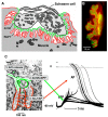
Postsynaptic folds and the anatomy of mAP generation. (A) Diagram of ion channel distribution at a rat NMJ. AChRs (green) are concentrated at the crests of the folds, while NaV1.4 channels are concentrated in the depths of the folds; (B) Fluorescence of rat NMJ showing AChRs (green) and NaV1.4 channels (red). Note that the NaV1.4 labeling protrudes beyond the AChRs because the NaV1.4 channels are located in the depths of the folds; (C) Anatomy of mAP generation. (1) ACh release from the nerve occurs next to the region of high AChR density (green); (2) This allows positive ions to enter the muscle, giving rise to the EPP; (3) The depolarization of the EPP opens the NaV1.4 channels in the depths of the folds, allowing a much bigger influx of positive ions which triggers that mAP. (From Slater, 2017, with permission) [21].
Figure 5
Figure 5
Patterns of vertebrate muscle innervation. Diagrams, based on nerve-specific stains, of the distribution of the motor nerve terminals in a range of vertebrate species. Note the variety of sizes and conformations, with human terminals among the smallest.
Figure 6
Figure 6
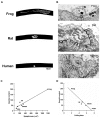
Correlations of structure and function in vertebrate NMJs. (A) Fluorescence images of frog, rat and human NMJs, labelled with α-bungarotoxin. Note that the human NMJ is much smaller than the other two; (B) EM images of NMJs in the same species. Note that the extent of postsynaptic folding increases from frog to human; (C) Quantal content is related to synaptic area in the vertebrate species studied; (D) Quantal content is related inversely to the extent of folding (folding index, FI).
Figure 7
Figure 7
Pathological features of NMJs. The images of mouse NMJs show, from left to right, the nerve, the distribution of AChRs and the ultrastructure. The upper images show NMJs from wild-type mice while the lower images show NMJs from dystrophin-deficient mdx mice, in which most muscle fibers degenerate, and then regenerate. Note the dramatic change in the conformation of the nerve terminal and the associated AChRs in the mdx mice, reflecting a new pattern of innervation established when the nerve reinnervates regenerated muscle fibers. In the EM images, note the presence of region of normal folding, presumed to be original contacts, adjacent to a region devoid of folding, presumed to be a new contact. Scale bars, light-micrographs, 20 μm, EM images, 1 μm. (From Lyons and Slater, 1991, with permission) [39]. The images of human NMJs are from a normal patient (top) and a patient with inherited AChR deficiency (bottom). Scale bar, 20 μm. (From Slater et al., 1997, with permission) [37].
Figure 8
Figure 8
Laminins at the mammalian NMJ. Diagrams of the distribution of various laminin forms at normal and abnormal mouse NMJs. (A) Wild type; (B) Loss of laminin β2; (C) Loss of laminin α2. Note that in the absence of laminin β2, there are almost no folds and the Schwann cell extends into the synaptic cleft. The loss of laminin α2 causes similar, but less dramatic changes. (Adapted from Rogers and Nishimune, 2016, with permission) [67].

References

    1. McMacken G., Abicht A., Evangelista T., Spendiff S., Lochmuller H. The Increasing Genetic and Phenotypical Diversity of Congenital Myasthenic Syndromes. Neuropediatrics. 2017;48:294–308. doi: 10.1055/s-0037-1602832. - DOI - PubMed
    1. Salpeter M.M. The Vertebrate Neuromuscular Junction. Alan R. Liss, Inc.; New York, NY, USA: 1986.
    1. Wu H., Xiong W.C., Mei L. To build a synapse: Signaling pathways in neuromuscular junction assembly. Development. 2010;137:1017–1033. doi: 10.1242/dev.038711. - DOI - PMC - PubMed
    1. Heuser J.E., Salpeter S.R. Organization of acetylcholine receptors in quick-frozen, deep-etched, and rotary-replicated Torpedo postsynaptic membrane. J. Cell Biol. 1979;82:150–173. doi: 10.1083/jcb.82.1.150. - DOI - PMC - PubMed
    1. Elmqvist D., Hofmann W.W., Kugelberg J., Quastel D.M.J. An electrophysiological investigation of neuromuscular transmission in myasthenia gravis. J. Physiol. 1964;174:417–434. doi: 10.1113/jphysiol.1964.sp007495. - DOI - PMC - PubMed

MeSH terms

LinkOut - more resources