Review
. 2017 Nov 1;41(6):723-750.
doi: 10.1093/femsre/fux048.

The role of toxins in Clostridium difficile infection

Affiliations
Review

The role of toxins in Clostridium difficile infection

Ramyavardhanee Chandrasekaran et al. FEMS Microbiol Rev. .

Abstract

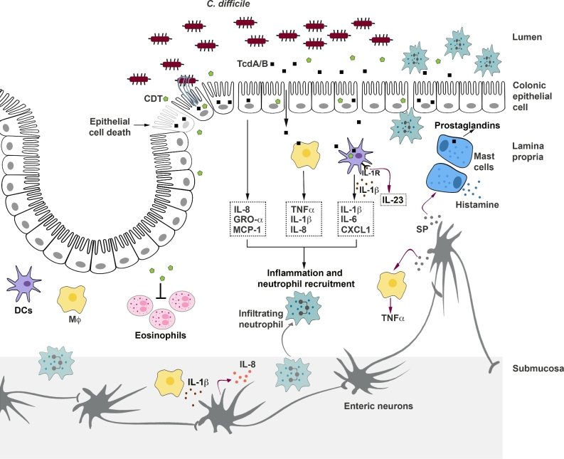
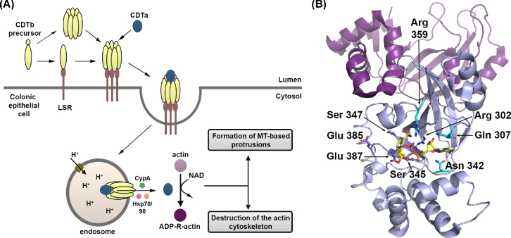
Clostridium difficile is a bacterial pathogen that is the leading cause of nosocomial antibiotic-associated diarrhea and pseudomembranous colitis worldwide. The incidence, severity, mortality and healthcare costs associated with C. difficile infection (CDI) are rising, making C. difficile a major threat to public health. Traditional treatments for CDI involve use of antibiotics such as metronidazole and vancomycin, but disease recurrence occurs in about 30% of patients, highlighting the need for new therapies. The pathogenesis of C. difficile is primarily mediated by the actions of two large clostridial glucosylating toxins, toxin A (TcdA) and toxin B (TcdB). Some strains produce a third toxin, the binary toxin C. difficile transferase, which can also contribute to C. difficile virulence and disease. These toxins act on the colonic epithelium and immune cells and induce a complex cascade of cellular events that result in fluid secretion, inflammation and tissue damage, which are the hallmark features of the disease. In this review, we summarize our current understanding of the structure and mechanism of action of the C. difficile toxins and their role in disease.

Keywords: Clostridium difficile; actin cytoskeleton; bacterial toxins; colitis; glucosyltransferase; inflammation; intestinal epithelium; pore formation.


Figures

Figure. 1.
Figure. 1.
Organization of toxin genes. (A) Schematic representation of the pathogenicity locus (PaLoc). Toxin-encoding genes, tcdA and tcdB, are indicated by blue arrows; regulatory genes are shown in light green (tcdR; positive) or red (tcdC; negative); and holin-encoding gene tcdE is shown in dark green. The direction of the arrows reflects the direction of transcription. TcdR positively regulates its own expression as well as the expression of tcdA and tcdB (indicated by brown arrows). TcdC is an anti-sigma factor that negatively regulates toxin expression by interfering with TcdR function. TcdE is involved in the secretion of toxins. (B) Schematic representation of the binary toxin locus (CdtLoc). CDT-encoding genes, cdtA and cdtB, are shown in blue. The regulatory gene cdtR is shown in light green. CdtR positively regulates the transcription of cdtA and cdtB.
Figure. 2.
Figure. 2.
TcdA and TcdB primary structure and mechanism of action. (A) TcdA and TcdB are organized into four functional domains: the glycosyltransferase domain (GTD; pink), the autoprocessing domain (APD; green), the delivery or pore-forming domain (blue) and the combined repetitive oligopeptides domain (CROPS; yellow). (B) The four functional domains contribute to a multistep mechanism of intoxication. TcdA and TcdB bind different cell surface proteins or sugars on the colonic epithelium (step 1) and are internalized by distinct endocytic pathways (step 2). The toxins reach acidified endosomes (step 3) and the low pH triggers a conformational change in the toxin delivery domain, resulting in pore formation and translocation of the GTD (and likely the APD) into the cytosol (step 4). Inositol hexakisphosphate (InsP6) binds and activates the APD, resulting in the cleavage and release of the GTD (step 5). The GTD inactivates Rho family proteins by transferring the glucose moiety (orange squares) from UDP-glucose to the switch I region of the GTPase (step 6). Glucosylation disrupts GTPase signaling and leads to cytopathic ‘rounding’ effects and apoptotic cell death.
Figure. 3.
Figure. 3.
Structure of the CROPS domain. (A) The CROPS domains of TcdA and TcdB consist of a series of short repeat (SR, yellow) sequences with interspersed long repeat (LR, purple) sequences. (B) A model of the full TcdA CROPS based on a fragment structure (2F6E) is corroborated by (C) negative stain electron microscopy images of TcdA (left) and TcdB (right). (D) The crystal structure of a CROPS fragment from the TcdA C-terminus (2G7C) shows how trisaccharides (orange carbons) bind at the vertices created at the intersection of an SR and LR.
Figure. 4.
Figure. 4.
TcdA structure. (A) The crystal structure of residues 4–1802 from TcdA (4R04) with the glucosyltransferase domain in pink and the autoprocessing domain in light green with its zinc atom depicted as an orange sphere. The delivery domain begins with a globular subdomain (sky blue), followed by an extended stretch of four hydrophobic α-helices (magenta). The α-helical stretch is scaffolded by an extended array of β-sheets (yellow). (B) The TcdA4-1802 structure docked into a 3D structure of the TcdA holotoxin obtained by negative stain electron microscopy (EM) at neutral pH. (C) The TcdA4-1802 structure docked into a 3D structure of the TcdA holotoxin obtained by negative stain EM at acidic pH indicates flexibility around the junction with the C-terminal CROPS domain.
Figure. 5.
Figure. 5.
The autoprocessing domain (APD) undergoes a significant conformational change upon binding to InsP6. The crystal structures of the TcdA APD in the (A) absence (4R04) and (B) presence of InsP6 (3HO6) reveal significant changes in the central β-flap structure (blue) and the C-terminal sequence that follows (teal). The structure of the APD in the context of TcdA4-1802 revealed the unexpected requirement for zinc (orange sphere) in TcdA and TcdB autoprocessing activity. Other key residues include Asp 589, His 655 and Cys 700 (side chains depicted with orange carbon atoms). His 759 is located at the tip of the β-flap and is bound to the zinc in the absence of InsP6. It moves significantly upon InsP6 binding.
Figure. 6.
Figure. 6.
The glucosyltransferase domain. The crystal structure of the TcdB glucosyltransferase domain (2BVL) with hydrolyzed UDP-glucose (yellow carbon atoms) bound in the core GT-A fold (light pink). The side chains of key catalytic residues (Trp 102, Asp 286, Asp 288 and Trp 520; aqua carbon atoms) are indicated along with the manganese atom (orange sphere). Residues that have been implicated in GTPase substrate recognition include Glu 449, Arg 455, Asp 461, Lys 463 and Glu 472 (green carbon atoms). The membrane localization domain corresponds to the four α-helices at the base of the structure (in purple) with the Phe 17 and Arg 18 residues implicated in membrane binding indicated with blue carbon atoms.
Figure. 7.
Figure. 7.
Cellular effects of C. difficile toxins. The toxins act on colonic epithelial cells and immune cells to induce inflammation and tissue damage. TcdA and TcdB disrupt the tight junctions and induce epithelial cell death, causing direct damage to the colonic epithelium. Additionally, the toxins stimulate epithelial cells to release inflammatory mediators that recruit neutrophils to the colonic mucosa. TcdA and TcdB can also enter the lamina propria following the disruption of the epithelial barrier and directly stimulate macrophages, dendritic cells, and mast cells to release inflammatory mediators, which further contribute to inflammation and neutrophil recruitment. Intoxication also results in the activation of enteric neurons and increased production of substance P (SP). SP can induce mast cell degranulation and can stimulate the lamina propria macrophages to release inflammatory cytokines. Prolonged intestinal inflammation can amplify tissue damage and contribute to neutrophil infiltration into the lumen, a key clinical feature of pseudomembranous colitis. The binary toxin CDT, expressed by some C. difficile strains, also induces cytopathic effects that lead to disruption of the tight junctions. Additionally, CDT can suppress a protective host eosinophilic response in the colon and can act synergistically with TcdA and TcdB to increase proinflammatory cytokine production by innate immune cells. Finally, CDT also contributes to C. difficile virulence by inducing the formation of microtubule-based cell protrusions that increase adherence of the bacteria.
Figure. 8.
Figure. 8.
TcdB-induced necrosis. At higher concentrations (100 pM and above), TcdB causes a necrotic form of cell death that does not require the autoprocessing and glucosyltransferase activities of the toxin. TcdB induces necrosis by promoting the assembly of the NADPH oxidase (NOX) complex on endosomes (step 1). The fully assembled NOX complex in the redox-active endosome mediates the transfer of an electron from NADPH to molecular oxygen, resulting in the generation of superoxide (step 2) and production of reactive oxygen species (ROS) (step 3). High levels of ROS promote cellular necrosis likely though DNA damage, lipid peroxidation, protein oxidation and/or mitochondrial dysfunction.
Figure. 9.
Figure. 9.
The CDT binary toxin. (A) Schematic representation of the mechanism of action of CDT. CDT consists of an enzymatic component (CDTa; blue) and a transport component (CDTb; yellow). The monomeric form of CDTb binds to its cell surface receptor, the lipolysis-stimulated lipoprotein receptor (LSR). Thereafter, CDTb undergoes proteolytic activation and oligomerization on the cell surface. Alternatively, the monomeric form of CDTb may undergo proteolytic activation and oligomer formation before binding to LSR. The oligomeric prepore binds CDTa, and the receptor-CDT complex is subsequently internalized into cells. As the endosome matures, the acidic pH triggers conformational changes in CDTb, resulting in membrane insertion and formation of a pore through which CDTa is translocated into the cytosol. Translocation of CDTa is facilitated by heat shock protein 70 (Hsp70), Hsp90 and cyclophilin A (CypA). In the cytosol, CDTa ADP-ribosylates actin at arginine-177. ADP-ribosylation of actin results in the eventual breakdown of the actin cytoskeleton and cytopathic cell rounding, and the formation of microtubule-based protrusions that enhance bacterial adherence. (B) The structure of the CDTa ADP-ribosyltransferase (2WN7). The N-terminal domain (purple) is responsible for binding CDTb, while the C-terminal domain (light blue) is responsible for the enzymatic activity. The side chains of residues within the RSE motif are depicted with blue carbons and are important for catalysis. Other residues important for binding of NAD (yellow carbons) are depicted with aqua carbons.

References

    1. Aktories K, Barbieri JT. Bacterial cytotoxins: targeting eukaryotic switches. Nat Rev Microbiol 2005;3:397–410. - PubMed
    1. Aktories K, Barmann M, Ohishi I et al. Botulinum C2 toxin ADP-ribosylates actin. Nature 1986;322:390–2. - PubMed
    1. Alcantara C, Stenson WF, Steiner TS et al. Role of inducible cyclooxygenase and prostaglandins in Clostridium difficile toxin A-induced secretion and inflammation in an animal model. J Infect Dis 2001;184:648–52. - PubMed
    1. Aldape MJ, Packham AE, Nute DW et al. Effects of ciprofloxacin on the expression and production of exotoxins by Clostridium difficile. J Med Microbiol 2013;62:741–7. - PMC - PubMed
    1. Alvin JW, Lacy DB. Clostridium difficile toxin glucosyltransferase domains in complex with a non-hydrolyzable UDP-glucose analogue. J Struct Biol 2017;198:203–9. - PMC - PubMed

MeSH terms

Substances