. 2018 Jan 1;35(1):132-148.
doi: 10.1093/molbev/msx274.

Linkage of A-to-I RNA Editing in Metazoans and the Impact on Genome Evolution

Affiliations

Linkage of A-to-I RNA Editing in Metazoans and the Impact on Genome Evolution

Yuange Duan et al. Mol Biol Evol. .

Abstract

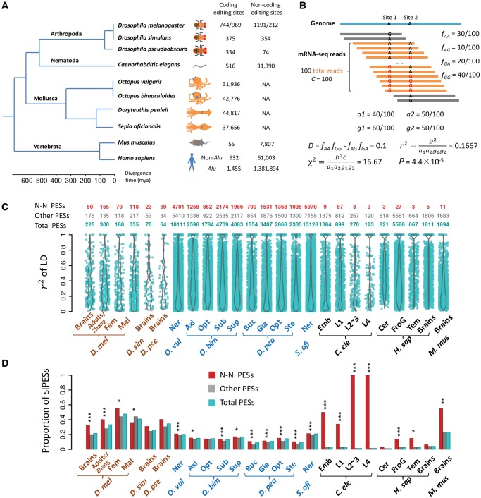
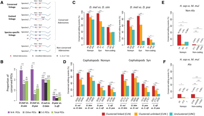
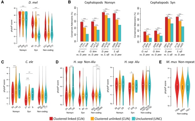
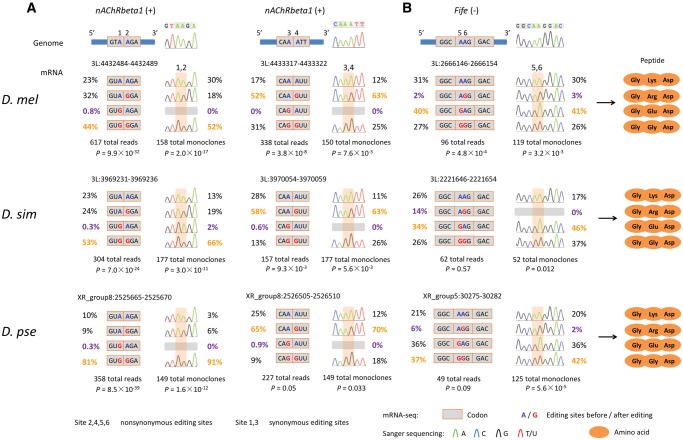
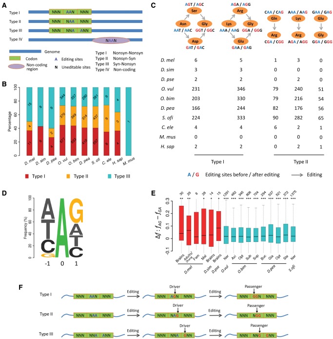
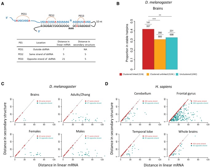
The adenosine-to-inosine (A-to-I) RNA editomes have been systematically characterized in various metazoan species, and many editing sites were found in clusters. However, it remains unclear whether the clustered editing sites tend to be linked in the same RNA molecules or not. By adopting a method originally designed to detect linkage disequilibrium of DNA mutations, we examined the editomes of ten metazoan species and detected extensive linkage of editing in Drosophila and cephalopods. The prevalent linkages of editing in these two clades, many of which are conserved between closely related species and might be associated with the adaptive proteomic recoding, are maintained by natural selection at the cost of genome evolution. Nevertheless, in worms and humans, we only detected modest proportions of linked editing events, the majority of which were not conserved. Furthermore, the linkage of editing in coding regions of worms and humans might be overall deleterious, which drives the evolution of DNA sites to escape promiscuous editing. Altogether, our results suggest that the linkage landscape of A-to-I editing has evolved during metazoan evolution. This present study also suggests that linkage of editing should be considered in elucidating the functional consequences of RNA editing.

Keywords: Drosophila; RNA editing; adaptive evolution; cephalopods; humans; linkage; mice; worms.


Figures

<sc>Fig</sc>. 1.
Fig. 1.
The landscape of linkage between RNA editing events in ten metazoan species. (A) A phylogenetic tree of the ten species we used in this study. The numbers of editing sites in coding and noncoding regions are given next to each species. For Drosophila melanogaster, the editing sites before and after the slash were retrieved from brains (Duan et al. 2017) and adults (Zhang et al. 2017), respectively. (B) A hypothetical example of calculating r2 between two editing sites based on the coverage of the NGS reads. Only reads covering the two editing sites (in orange) were included, and the reads spanning only one editing site (in gray) were discarded. (C) Violin plots of r2 (y-axis) for significantly linked PESs in 25 samples of ten species (x-axis). The numbers of total, N–N and the remaining slPESs in each sample are presented above the plots. (D) The proportions of the PESs that have editing events significantly linked in each sample (adjusted P < 0.05). Besides the total PESs, the fraction for the N–N or the remaining PESs that was significantly linked was also calculated in each sample. The fraction of the N–N PESs that is significantly linked was compared with that of the remaining PESs with the Fisher’s exact test. *P < 0.05; **P < 0.01; ***P < 0.001. For the four species O. bimaculoides, D. pealeii, C. elegans, and H. sapiens, only the top four samples with the largest numbers of slPESs are shown in (C) and (D). Fem, female adults; Mal, male adults; Ner, nerve system; Axi, axial nerve cord; Opt, Optic lobe; Sub, subesophageal brain; Sup, supraesophageal brain; Buc, Buccal ganglia; Gia, giant fiber lobe; Ste, stellate ganglion; Emb, embryos; L1, L1 larvae; L2∼3, L2∼3 larvae; L4, L4 larvae; Cer, cerebellum; FroG, frontal gyrus; Tem, temporal lobe. D. mel, Drosophila melanogaster; D. sim, Drosophila simulans; D. pse, Drosophila pseudoobscura; O. vul, Octopus vulgaris; O. bim, Octopus bimaculoides; D. pea, Doryteuthis pealeii; S. ofi, Sepia oficianalis; C. ele, Caenorhabditis elegans; H. sap, Homo sapiens; M. mus, Mus musculus.
<sc>Fig</sc>. 2.
Fig. 2.
Conservation of linked editing events between species. (A) The possible conservation patterns of a slPES in another species: 1) conserved linkage, for which the two orthologous adenosine sites are both edited and significantly linked in another species; 2) evolved linkage, for which one of the orthologous adenosine sites is edited but linked with a different editing site; 3) species-specific linkage, for which the two orthologous sites might be edited but do not show linkage to each other, and they are not linked with other editing sites as well. It is also possible that the orthologous sites in another species are not adenosines (B, which represents C, T, or G) so that editing would not occur on the orthologous sites. (B) The proportion (y-axis) of the total, the N–N, and the remaining slPESs that are conserved between two Drosophila species or between two cephalopod species (***P < 0.001; Fisher’s exact tests). In each comparison, the numbers of the tested slPESs and the conserved slPESs between species are given above the plot. In flies, all the remaining (Other) slPESs were used to compare with the N–N slPESs to increase the statistical power; and in cephalopods, the S–S (synonymous–synonymous) slPESs were used to compare with the N–N slPESs. (CF) The proportions of the editing events that are evolutionarily conserved between two species of Drosophila (brains, C), cephalopods (pooled tissues, D), non-Alu (E) and Alu (F) regions between humans and rhesus macaque (prefrontal cortex and cerebellum). Nonsynonymous (Nonsyn), synonymous (Syn) and noncoding adenosine sites are divided into three categories: 1) clustered (within 100 nt) and significantly linked (CLN); 2) clustered but unlinked (CUN); and 3) unclustered (UNC). When comparing editing events in two species, only the sites with editing level ≥ 0.05 and the genomic DNA were adenosines in both species were considered. The numbers of total editing sites in each category used for comparison are presented below the bars. The Fisher’s exact tests were performed to detect statistical significance (*P < 0.05; **P < 0.01; ***P < 0.001).
<sc>Fig</sc>. 3.
Fig. 3.
Conservation patterns of the CLN, CUN, and UNC editing sites at DNA level. (A) The violin plots of phyloP scores (y-axis) of the CLN, CUN, and UNC editing sites in the nonsynonymous (Nonsyn), synonymous (Syn) and noncoding functional categories in D. melanogaster. (B) The proportions of adenosine sites that were edited in a cephalopod species (the former in a comparison) and have orthologous sites to be adenosines in another cephalopod species (the latter in a comparison). The Fisher’s exact test was performed to test differences in the proportions of conserved adenosine sites (*P < 0.05; **P < 0.01; ***P < 0.001). (CE) The violin plots of phyloP scores (y-axis) of the CLN, CUN, and UNC editing sites in the Nonsyn, Syn and noncoding editing functional categories in C. elegans (C), non-Alu and Alu regions of humans (D) and nonrepetitive regions of mice (E). The numbers of total editing sites in each category are given below the violins or bars. For flies, worms, humans, and mice, only editing sites with phyloP scores available are considered and the Wilcoxon rank-sum test was performed to test differences in phyloP scores (*P < 0.05; **P < 0.01; ***P < 0.001).
<sc>Fig</sc>. 4.
Fig. 4.
Sanger verification of linked editing events in three Drosophila species. (A) In nAChRbeta1, two pairs of adjacent editing sites (sites 1 and 2, and sites 3 and 4) are highly linked in brains of all the three Drosophila species. For each of the two PESs, the first editing site is located at the third base of a codon, and the other linked editing site is located at the first base of next codon. (B) In Fife, a linked pair of editing sites (sites 5 and 6) that are located at the first and second bases of the same codon, is conserved across three Drosophila species. The linkage events revealed by NGS reads were verified by the Sanger sequencing of cDNA monoclones. For each of the three double editing pairs, the genomic location, haplotype frequencies, the resultant amino acids, and the depth of the NGS reads and Sanger sequencing results, as well as examples of Sanger sequencing traces, are shown.
<sc>Fig</sc>. 5.
Fig. 5.
The nonindependent editing of the AA dinucleotides and the implications for functional prediction. (A) Four types of AA double editing sites that are significantly linked. The first three types are located in CDSs, and the fourth type is in the noncoding region. (B) Numbers (in each bar) and percentages (y-axis) of the three types of AA double editing sites that are significantly linked (adjusted P < 0.05) in each species. (C) Summary of the type I and type II PESs that are significantly linked in the pooled samples of each species. The codons containing type I editing sites originally encode Asn (AAT or AAC) or Lys (AAA or AAG). When both adenosines are edited, the encoded amino acid becomes Gly (GGA, GGC, GGG, or GGT). For codons containing type II editing sites, the amino acids encoded are merely decided by whether the front editing sites are edited or not. (D) The triplet centered with the focal editing sites based on our previous study (Duan et al. 2017). (E) The differences in frequency (Δf) of “AG” (fAG) and “GA” (fGA) haplotypes among all the significantly linked editing events in the AA dinucleotides in CDSs of 16 representative samples. In each sample, the differences in frequency of two haplotypes were compared with the Wilcoxon sign-rank test (*P < 0.05; **P < 0.01; ***P < 0.001). (F) The nonindependent editing of the AA dinucleotides. Editing of the rear adenosine (Driver) facilities editing of the front adenosine (Passenger) in an AA dinucleotide.
<sc>Fig</sc>. 6.
Fig. 6.
The spatial distance of two linked editing sites in a hairpin structure. (A) The scheme illustrating the distance between two linked editing sites in a hairpin structure. Examples of PESs that are outside the hairpin (PES1), in the same (PES2) or opposite sides (PES3) of a stable hairpin structure. The distances between two editing sites in the linear mRNA and the secondary structure are show in the table below. The distance in the secondary structure was calculated as the length of the shortest path between two editing sites by treating the hairpin as a graph. (B) The proportion of editing sites in brains of D. melanogaster that are located in the stable hairpins. The editing sites were divided into CLN, CUN, and UNC categories. In each category, the numbers of total editing sites and those in stable hairpins are given above the bars. Fisher’s exact tests were performed to detect the statistical differences (**P < 0.01; ***P < 0.001). (C) The linear (x-axis) and spatial (y-axis) distance (nt) between the significantly linked editing sites that were located in the same (red) or opposite (cyan) sides of hairpin structures in four samples of D. melanogaster. (D) Similar as (C) but shows the results for four human brain samples.

Similar articles

Cited by

References

    1. Alon S, Garrett SC, Levanon EY, Olson S, Graveley BR, Rosenthal JJC, Eisenberg E.. 2015. The majority of transcripts in the squid nervous system are extensively recoded by A-to-I RNA editing. eLife 4:e05198. - PMC - PubMed
    1. Alon S, Mor E, Vigneault F, Church GM, Locatelli F, Galeano F, Gallo A, Shomron N, Eisenberg E.. 2012. Systematic identification of edited microRNAs in the human brain. Genome Res. 22(8): 1533–1540. - PMC - PubMed
    1. Athanasiadis A, Rich A, Maas S.. 2004. Widespread A-to-I RNA editing of Alu-containing mRNAs in the human transcriptome. PLoS Biol. 2(12): e391.. - PMC - PubMed
    1. Barrett JC, Fry B, Maller J, Daly MJ.. 2005. Haploview: analysis and visualization of LD and haplotype maps. Bioinformatics 21(2): 263–265.http://dx.doi.org/10.1093/bioinformatics/bth457 - DOI - PubMed
    1. Bass BL. 2002. RNA editing by adenosine deaminases that act on RNA. Annu Rev Biochem. 71:817–846.http://dx.doi.org/10.1146/annurev.biochem.71.110601.135501 - DOI - PMC - PubMed

Publication types

MeSH terms

LinkOut - more resources