. 2017 Nov;51(5):1439-1448.
doi: 10.3892/ijo.2017.4132. Epub 2017 Sep 22.

Brain lipid-binding protein promotes proliferation and modulates cell cycle in C6 rat glioma cells

Affiliations

Brain lipid-binding protein promotes proliferation and modulates cell cycle in C6 rat glioma cells

Xiao Han et al. Int J Oncol. 2017 Nov.

Abstract

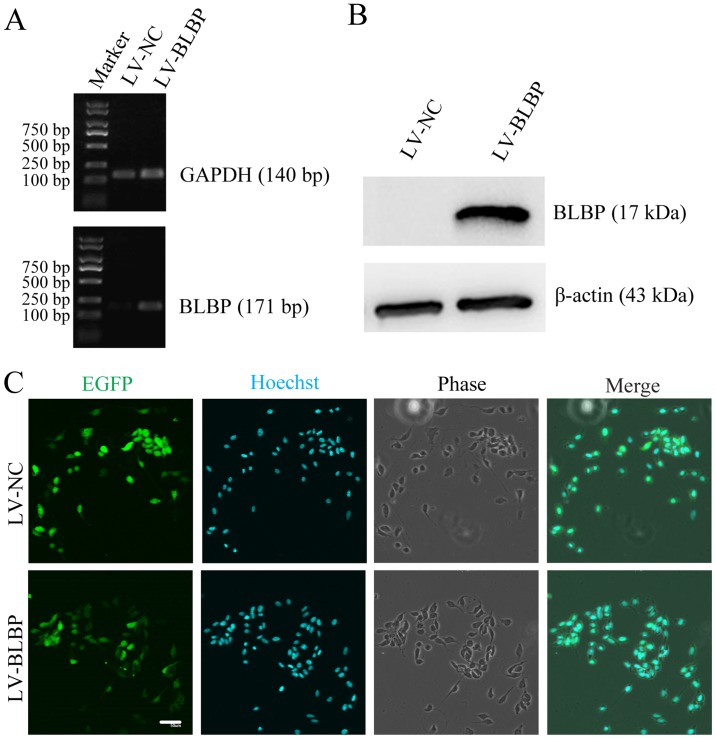
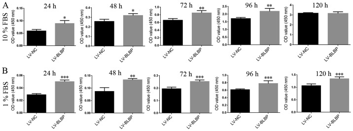
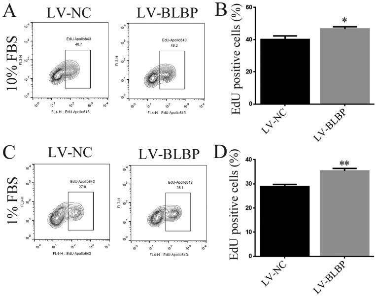
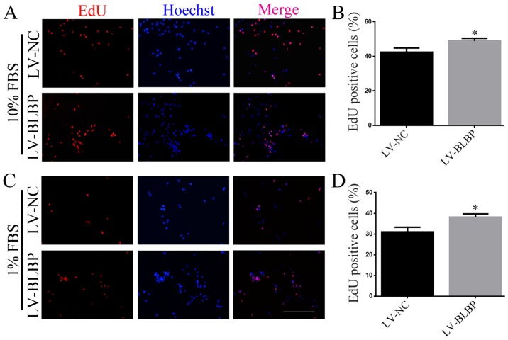
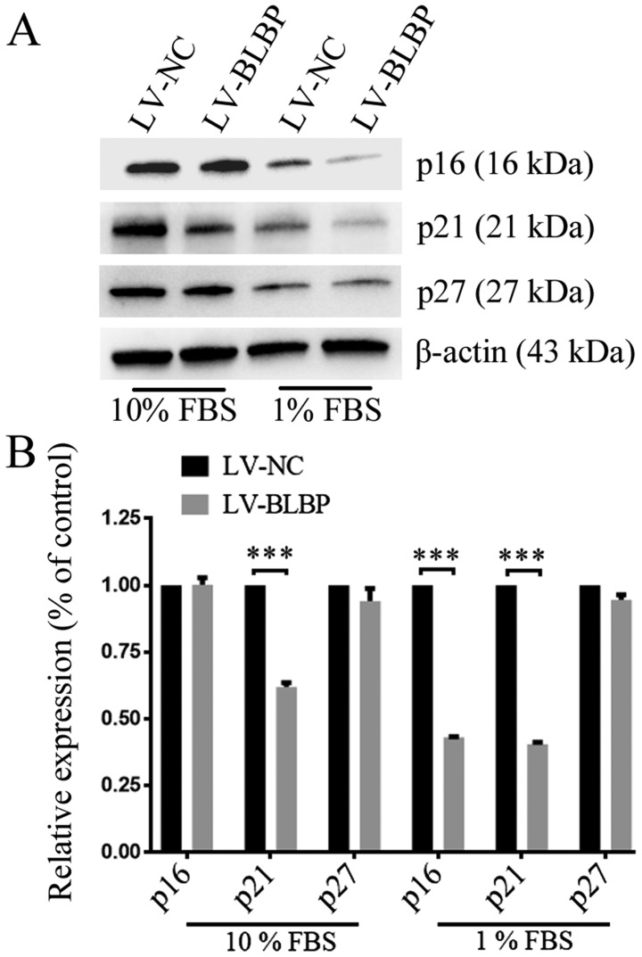
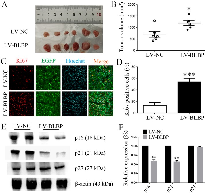
Gliomas are the most common primary brain tumors affecting adults. Four grades of gliomas have been identified, namely, grades I-IV. Brain lipid-binding protein (BLBP), which functions in the intracellular transport of fatty acids, is expressed in all grades of human gliomas. The glioma cells that are cultured in vitro are grouped into the BLBP-positive and BLBP-negative cell lines. In the present study, we found that C6 rat glioma cells was a distinct type of BLBP-negative cell line. Our results confirmed that in the C6 cells, the expression of exogenous BLBP increased the proliferation and percentage of cells in the S phase, in the culture medium containing 10 or 1% FBS. Moreover, exogenous BLBP was found to downregulate the tumor suppressors p21 and p16 in the 1% FBS culture medium, but only p21 in the 10% FBS culture medium. The results of the xenograft model assay showed that exogenous BLBP also stimulated tumor formation and downregulated p21 and p16. In conclusion, our study demonstrated that exogenous BLBP promoted proliferation of the C6 cells in vitro and facilitated tumor formation in vivo. Therefore, BLBP expression in glioma cells may promote cell growth by inhibiting the tumor suppressors.


Figures

Figure 1
Figure 1
Following infection with lentivirus, the PCR results showed that the BLBP gene was positively amplified in the cells from the LV-BLBP group but not in those from the LV-NC group (A), and BLBP protein was detected only in the cells from the LV-BLBP group (B); all C6 cells both in LV-NC (C, upper panel) and LV-BLBP (C, lower panel) groups expressed EGFP, scale bar=50 µm.
Figure 2
Figure 2
Cell viability of C6 cells was determined using the CCK-8 reagent by measuring the optical density (OD) values at 450 nm. When cultured in the 10% FBS medium, the OD values of cells from the LV-BLBP group increased from 24 to 96 h (*P<0.05; **P<0.01), but not at 120 h. After culturing in the 1% FBS medium, the OD values of cells from the LV-BLBP group were elevated from 24 to 120 h (**P<0.01; ***P<0.001).
Figure 3
Figure 3
FACS analysis showed that the proportion of cells in the S phase increased (C and F) in the LV-BLBP group, both in the 10% (A and B) and 1% FBS (D and e) culture medium (**P<0.01); however, the proportion of cells in the G0/G1 phase decreased (***P<0.001).
Figure 4
Figure 4
The EdU-labeled cells cultured in the 10% (A and B) or 1% FBS (C and D) medium were analyzed using FACS. Results showed that cells from the LV-BLBP group had a significantly higher number of EdU-labeled cells than those from the LV-NC group (*P<0.05, **P<0.01).
Figure 5
Figure 5
EdU-positive cells were examined under the microscope. The number of EdU-positive cells was higher in the LV-BLBP group in both the 10% FBS (A and B) and 1% FBS (C and D) culture medium (*P<0.05); scale bar, 200 µm.
Figure 6
Figure 6
In western blot assay (A), both p21 and p16 expression levels decreased in cells in the LV-BLBP group when cultured in 1% FBS medium. However, only p21 levels decreased in cells in the LV-BLBP group when cultured in 10% FBS medium. No differences in p27 levels were observed between the two groups either in the 10% FBS or 1% FBS culture medium (B) (***P<0.001).
Figure 7
Figure 7
Tumor formation assay results showed that C6 cells from both the LV-NC group (A, upper panel) and LV-BLBP group (A, lower panel) formed tumors after subcutaneous injection for 21 days. Tumor volumes in mice from LV-BLBP were larger than those in the LV-NC group (B) (*P<0.05). The number of Ki67-positive cells (C) in tumor tissues of mice from the LV-BLBP group were considerably higher than those in the LV-NC group (D, ***P<0.001), scale bar, 50 µm. expression levels of p21 and p16, but not p27, were markedly lower in the tumor tissues of mice in the LV-BLBP group (E and F) (**P<0.01).
Figure 8
Figure 8
In the 10% FBS medium (A), cells from the LV-NC and LV-BLBP groups show no significant differences in cell migration distances at 12 h (B), scale bar, 400 µm. In the 1% FBS medium (C), cells from the LV-NC and LV-BLBP groups showed no significant differences in cell migration distances at 12, 24, and 48 h (D), scale bar, 400 µm.

Similar articles

Cited by

References

    1. Furnari FB, Fenton T, Bachoo RM, Mukasa A, Stommel JM, Stegh A, Hahn WC, Ligon KL, Louis DN, Brennan C, et al. Malignant astrocytic glioma: Genetics, biology, and paths to treatment. Genes Dev. 2007;21:2683–2710. doi: 10.1101/gad.1596707. - DOI - PubMed
    1. Rich JN, Eyler CE. Cancer stem cells in brain tumor biology. Cold Spring Harb Symp Quant Biol. 2008;73:411–420. doi: 10.1101/sqb.2008.73.060. - DOI - PMC - PubMed
    1. Louis DN, Ohgaki H, Wiestler OD, Cavenee WK, Burger PC, Jouvet A, Scheithauer BW, Kleihues P. The 2007 WHO classification of tumours of the central nervous system. Acta Neuropathol. 2007;114:97–109. doi: 10.1007/s00401-007-0243-4. - DOI - PMC - PubMed
    1. Balendiran GK, Schnutgen F, Scapin G, Borchers T, Xhong N, Lim K, Godbout R, Spener F, Sacchettini JC. Crystal structure and thermodynamic analysis of human brain fatty acid-binding protein. J Biol Chem. 2000;275:27045–27054. - PubMed
    1. Liang Y, Bollen AW, Aldape KD, Gupta N. Nuclear FABP7 immunoreactivity is preferentially expressed in infiltrative glioma and is associated with poor prognosis in EGFR-overexpressing glioblastoma. BMC Cancer. 2006;6:97. doi: 10.1186/1471-2407-6-97. - DOI - PMC - PubMed