. 2017 Nov;51(5):1601-1610.
doi: 10.3892/ijo.2017.4138. Epub 2017 Sep 27.

Dual action of NSC606985 on cell growth and apoptosis mediated through PKCδ in prostatic cancer cells

Affiliations

Dual action of NSC606985 on cell growth and apoptosis mediated through PKCδ in prostatic cancer cells

Xin Wang et al. Int J Oncol. 2017 Nov.

Abstract

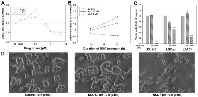
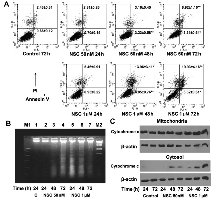
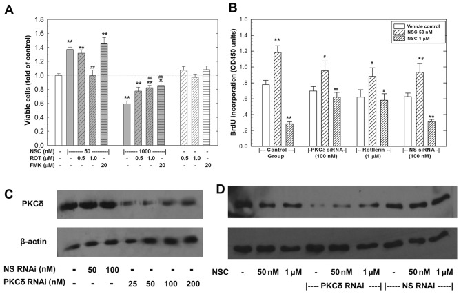
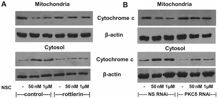
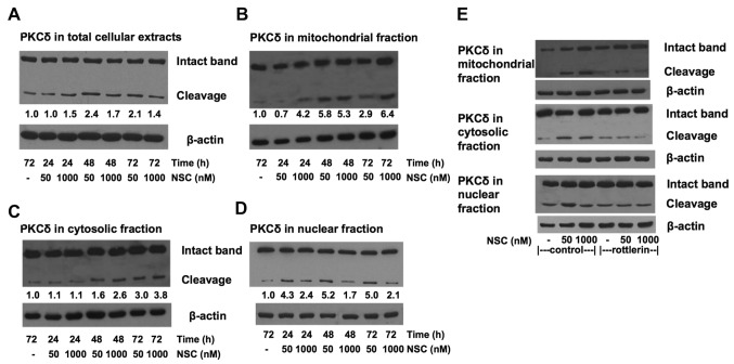
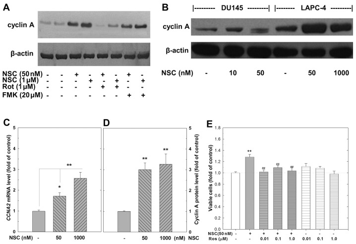
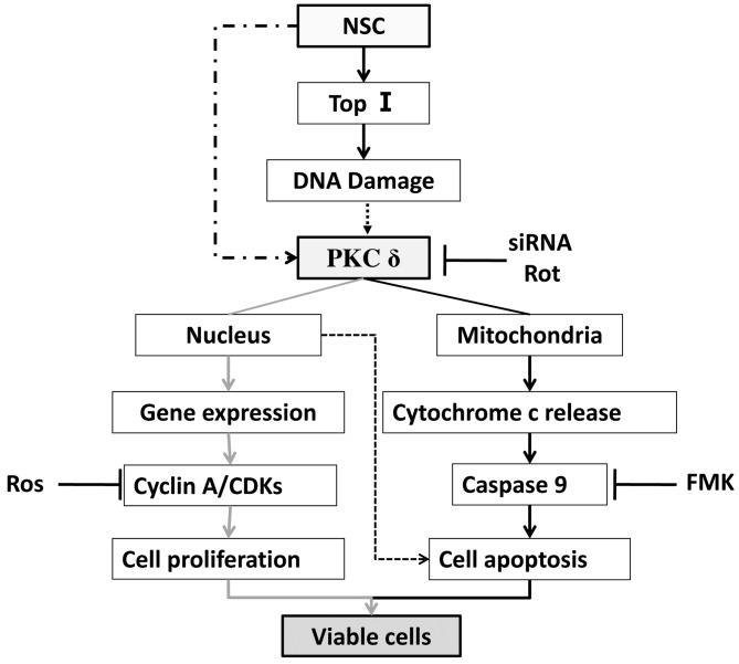
Chemotherapy is a vital therapeutic strategy for castration-resistant prostate cancer (CRPC). We have previously shown that NSC606985 (NSC), a camptothecin (CPT) analog, induced cell apoptosis via interacting with topoisomerase I (Topo I) in prostate cancer cells. In the present study, the effect and mechanism of CPT analogs in LAPC4 cells were investigated. LAPC-4 cells were treated with NSC, CPT, and topotecan. Cell proliferation, apoptosis, and protein kinase Cδ (PKCδ) subcellular activation were measured at different doses and time-points, with or without PKCδ inhibition or knockdown of PKCδ expression. NSC at doses ranging from 10 to 100 nM induced a dose-dependent increase in viable cell number and DNA biosynthesis with mild cell apoptosis, whereas, at doses ranging from 500 nM to 5 mM, NSC produced a dose-dependent decrease in cell proliferation and DNA biosynthesis with a significant induction of cell apoptosis. Both NSC-induced cell proliferation and apoptosis were blocked by knockdown of PKCδ with a specific RNAi, or by the co-administration of rottlerin, a PKCδ inhibitor. Moreover, NSC produced a dose-dependent subcellular activation of PKCδ. The dose-dependent dual action of NSC is mediated at least in part through the differential subcellular activation of PKCδ in LAPC4 cells. The demonstration of a differential cell response to camptothecin analogs would facilitate the identification of biomarker(s) to CPT sensitivity and promote the personalization of CPT chemotherapy in CRPC.


Figures

Figure 1
Figure 1
CPT and its analogs (NSC and topotecan) produce biphasic effect on cell growth and death in PCa cells. (A) Dose-dependent dual action of NSC and CPT on viable cell number in LAPC4 cells after 72 h treatment. The data are shown as mean ± SEM, n=6–12 for NSC, n=3–6 for CPT. (B) Time- and dose-dependent dual action of NSC on viable cell number in LAPC4 cells. The data are shown as mean ± SEM, (n=3). (C) Effects of topotecan treatment for 72 h on cell viability in different PCa cell lines. The data are shown as mean ± SEM, n=6–9. (D) Morphological changes of LAPC4 cells after NSC treatment (72 h). *p<0.05 and **p<0.001 compared to corresponding controls. CPT, camptothecin.
Figure 2
Figure 2
NSC induces cell apoptosis in LAPC4 cells. (A) Time- and dose-dependent apoptosis of LAPC4 cells after NSC treatment. The data are shown as mean ± SEM, n=6. *p<0.05 and **p<0.01 compared to control. (B) NSC induced DNA fragmentation in LAPC4 cells. (C) NSC induced cytochrome c release from mitochondria to cytosol in LAPC4 cells.
Figure 3
Figure 3
The dual action of NSC involves PKCδ in LAPC4 cells. (A) Rottlerin (ROT) blocked NSC-induced biphasic effect in LAPC4 cells. (B) NSC-induced biphasic effect on DNA biosynthesis was blocked by rottlerin and knockdown of PKCδ in LAPC4 cells. The data in (A and B) are shown as mean ± SEM, n=6–9. *p<0.05 and **p<0.01 compared to control; ##p<0.01 compared to corresponding NSC treatment. (C and D) Confirmation of the knockdown of PKCδ by RNAi transfection with or without NSC treatment. ROT, rottlerin; FMK, Z-VAD-fluoromethylketone; NS RNAi, non-specific RNAi; PKCδ, protein kinase Cδ.
Figure 4
Figure 4
NSC-induced cytochrome c release from mitochondria to cytosol is blocked by rottlerin and knockdown of PKCδ in LAPC4 cells (72 h). (A) Cytochrome c release after 72 h NSC treatment with or without co-treatment of rottlerin (1 µM). (B) Cytochrome c release after 72 h NSC treatment with 24 h pre-transfection of either NS RNAi (100 nM), or PKCδ RNAi (100 nM). NS RNAi, non-specific RNAi. PKCδ, protein kinase Cδ.
Figure 5
Figure 5
NSC induces proteolytic cleavage of PKCδ in a subcellular compartment-specific manner in LAPC4 cells. (A) Total cellular PKCδ cleavage in LAPC4 cells after NSC treatment. (B) Mitochondrial PKCδ cleavage in LAPC4 cells after NSC treatment. (C) Cytosolic PKCδ cleavage in LAPC4 cells after NSC treatment. (D) Nuclear PKCδ cleavage in LAPC4 cells after NSC treatment. (E) Differential subsellular PKCδ cleavage in LAPC4 cells after NSC treatment with or without rottlerin (1 µM) co-treatment (72 h). The quantitative changes in PKCδ cleavage are indicated at the bottom of the PKCδ western blot analyses (A–D) expressed as folds of control. PKCδ, protein kinase Cδ.
Figure 6
Figure 6
NSC-induced cell growth involves upregulation of cyclin A expression in LAPC4 cells. (A and B) Cyclin A expression in LAPC4 and DU145 cells after 72 h treatment of various regiments as indicated. (C and D) CCNA2 mRNA and cyclin A protein expression in LAPC4 cells after 72 h NSC treatment, respectively. The data are shown as mean ± SEM, n=5–9. (E) Effects of roscovitine (Ros) on cell growth in NSC-treated (50 nM) LAPC4 cells (72 h). The data are shown as mean ± SEM, n=6. *p<0.05 and **p<0.01 compared to control; ##p<0.01 compared to NSC treatment alone. ROT, rottlerin; FMK, Z-VAD-fluoromethylketone; Ros, roscovitine.
Figure 7
Figure 7
A schematic representation of NSC-induced dual action on cell proliferation and apoptosis in LAPC4 cells. Solid lines indicate defined actions and dashed lines possible actions. Top I, topoisomerase I; Rot, rottlerin; Ros, roscovitine; FMK, Z-VAD-fluoromethylketone; CDKs, cyclin dependent kinases.

Similar articles

Cited by

References

    1. Siegel R, Ma J, Zou Z, Jemal A. Cancer statistics, 2014. CA Cancer J Clin. 2014;64:9–29. doi: 10.3322/caac.21208. - DOI - PubMed
    1. Petrylak DP. The treatment of hormone-refractory prostate cancer: Docetaxel and beyond. Rev Urol. 2006;8(Suppl 2):S48–S55. - PMC - PubMed
    1. Silvestris N, Leone B, Numico G, Lorusso V, De Lena M. Present status and perspectives in the treatment of hormone-refractory prostate cancer. Oncology. 2005;69:273–282. doi: 10.1159/000089676. - DOI - PubMed
    1. Aziz MH, Dreckschmidt NE, Verma AK. Plumbagin, a medicinal plant-derived naphthoquinone, is a novel inhibitor of the growth and invasion of hormone-refractory prostate cancer. Cancer Res. 2008;68:9024–9032. doi: 10.1158/0008-5472.CAN-08-2494. - DOI - PMC - PubMed
    1. Frese S, Schüller A, Frese-Schaper M, Gugger M, Schmid RA. Cytotoxic effects of camptothecin and cisplatin combined with tumor necrosis factor-related apoptosis-inducing ligand (Apo2L/TRAIL) in a model of primary culture of non-small cell lung cancer. Anticancer Res. 2009;29:2905–2911. - PubMed

MeSH terms