

Reversal effect of adenovirus-mediated human interleukin 24 transfection on the cisplatin resistance of A549/DDP lung cancer cells
- PMID: 29048638
- PMCID: PMC5780038
- DOI: 10.3892/or.2017.6002
Reversal effect of adenovirus-mediated human interleukin 24 transfection on the cisplatin resistance of A549/DDP lung cancer cells
Abstract
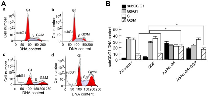
Interleukin-24 (IL-24) is a tumor-suppressor gene that has been documented in human melanoma cells. IL-24 has marked antitumor activities on various types of human cancer, but its underlying mechanism remains unclear. In the present, we investigated the effects of human IL-24 (hIL-24) on the chemotherapy resistance of lung cancer cells. The cisplatin (DDP)-resistant lung carcinoma cell line A549/DDP was subjected to adenovirus-mediated transfection with the human IL-24 gene (Ad-hIL-24). The growth-inhibitory and apoptotic effects of Ad-hIL-24 on A549/DDP cells were observed, and the expression levels of AKT, phosphorylated-AKT (p-AKT) and P-glycoprotein (P-gp) were detected. Ad-hIL-24 significantly decreased the levels of p-AKT and P-gp, and effectively inhibited A549/DDP cell growth. Furthermore, A549/DDP cells exhibited a significantly increased rate of apoptosis, as well as G2/M-phase arrest, following transfection with Ad-hIL-24, and these effects were increased in cells treated with Ad-IL-24 combined with DDP when compared with those treated with Ad-hIL-24 or DDP alone. These results suggest that hIL-24 can reverse the DDP resistance of lung cancer cells, and that the associated mechanism involves the induction of apoptosis and G2/M-phase arrest through the phosphoinositide3-kinase (PI3K)/AKT signaling pathway, as well as a decrease in drug resistance through P-gp expression.
Figures


Similar articles
-
Baicalin attenuates DDP (cisplatin) resistance in lung cancer by downregulating MARK2 and p-Akt.Int J Oncol. 2017 Jan;50(1):93-100. doi: 10.3892/ijo.2016.3768. Epub 2016 Nov 15. Int J Oncol. 2017. PMID: 27878245
-
Co-treatment with BEZ235 enhances chemosensitivity of A549/DDP cells to cisplatin via inhibition of PI3K/Akt/mTOR signaling and downregulation of ERCC1 expression.Oncol Rep. 2018 Oct;40(4):2353-2362. doi: 10.3892/or.2018.6583. Epub 2018 Jul 20. Oncol Rep. 2018. PMID: 30066933
-
XPC inhibition rescues cisplatin resistance via the Akt/mTOR signaling pathway in A549/DDP lung adenocarcinoma cells.Oncol Rep. 2019 Mar;41(3):1875-1882. doi: 10.3892/or.2019.6959. Epub 2019 Jan 9. Oncol Rep. 2019. PMID: 30628719
-
Silencing long non-coding RNA ROR improves sensitivity of non-small-cell lung cancer to cisplatin resistance by inhibiting PI3K/Akt/mTOR signaling pathway.Tumour Biol. 2017 May;39(5):1010428317697568. doi: 10.1177/1010428317697568. Tumour Biol. 2017. PMID: 28459375
-
Effects of VBMDMP on the reversal of cisplatin resistance in human lung cancer A549/DDP cells.Oncol Rep. 2015 Jan;33(1):372-82. doi: 10.3892/or.2014.3607. Epub 2014 Nov 13. Oncol Rep. 2015. PMID: 25394854
Cited by
-
WT1 Inhibits Human Renal Carcinoma Cell Proliferation and Induces G2/M Arrest by Upregulating IL-24 Expression.Biomed Res Int. 2022 Jul 23;2022:1093945. doi: 10.1155/2022/1093945. eCollection 2022. Biomed Res Int. 2022. PMID: 35915803 Free PMC article.
-
Adenovirus-Mediated LAMA3 Transduction Enhances Hemidesmosome Formation and Periodontal Reattachment during Wound Healing.Mol Ther Methods Clin Dev. 2020 Jun 4;18:291-303. doi: 10.1016/j.omtm.2020.06.001. eCollection 2020 Sep 11. Mol Ther Methods Clin Dev. 2020. PMID: 32671133 Free PMC article.
-
Nuclear factor-erythroid 2-related factor 3 (NRF3) is low expressed in colorectal cancer and its down-regulation promotes colorectal cancer malignance through activating EGFR and p38/MAPK.Am J Cancer Res. 2019 Mar 1;9(3):511-528. eCollection 2019. Am J Cancer Res. 2019. Retraction in: Am J Cancer Res. 2024 Aug 25;14(8):4112. doi: 10.62347/VVLN6538. PMID: 30949407 Free PMC article. Retracted.
-
Ad-VT enhances the sensitivity of chemotherapy-resistant lung adenocarcinoma cells to gemcitabine and paclitaxel in vitro and in vivo.Invest New Drugs. 2022 Apr;40(2):274-289. doi: 10.1007/s10637-021-01204-4. Epub 2022 Jan 4. Invest New Drugs. 2022. PMID: 34981275 Free PMC article.
-
Suppression of YAP by DDP disrupts colon tumor progression.Oncol Rep. 2018 May;39(5):2114-2126. doi: 10.3892/or.2018.6297. Epub 2018 Mar 6. Oncol Rep. 2018. Retraction in: Oncol Rep. 2021 Jan;45(1):406. doi: 10.3892/or.2020.7843. PMID: 29512779 Free PMC article. Retracted.
References
-
- Takase N, Hattori Y, Kiriu T, Itoh S, Kawa Y, Yamamoto M, Urata Y, Shimada T, Tsujino K, Soejima T, et al. Concurrent chemoradiotherapy with cisplatin and S-1 or vinorelbine for patients with stage III unresectable non-small cell lung cancer: A retrospective study. Respir Investig. 2016;54:334–340. doi: 10.1016/j.resinv.2016.02.008. - DOI - PubMed
-
- Lunacsek OE, Ravelo A, Coutinho AD, Hazard SJ, Green MR, Willey J, Eaddy M, Goertz HP. First-line treatment with bevacizumab and platinum doublet combination in non-squamous non-small cell lung cancer: A retrospective cohort study in US oncology community practices. Drugs Real World Outcomes. 2016;3:333–343. doi: 10.1007/s40801-016-0090-5. - DOI - PMC - PubMed
MeSH terms
Substances
LinkOut - more resources
Full Text Sources
Other Literature Sources
Medical
Miscellaneous
