. 2017 Oct 19;12(10):e0186900.
doi: 10.1371/journal.pone.0186900. eCollection 2017.

XRCC5 cooperates with p300 to promote cyclooxygenase-2 expression and tumor growth in colon cancers

Affiliations

XRCC5 cooperates with p300 to promote cyclooxygenase-2 expression and tumor growth in colon cancers

Zhifeng Zhang et al. PLoS One. .

Abstract

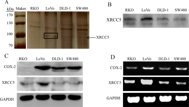
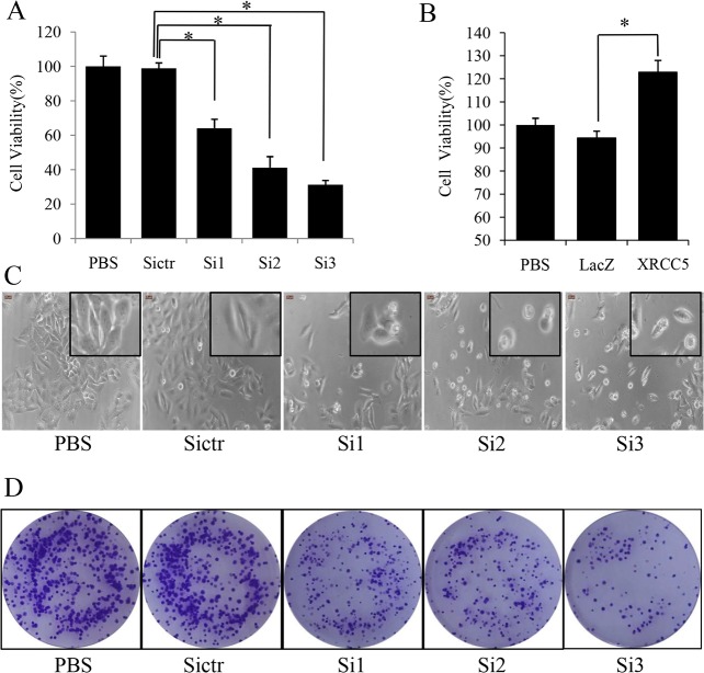
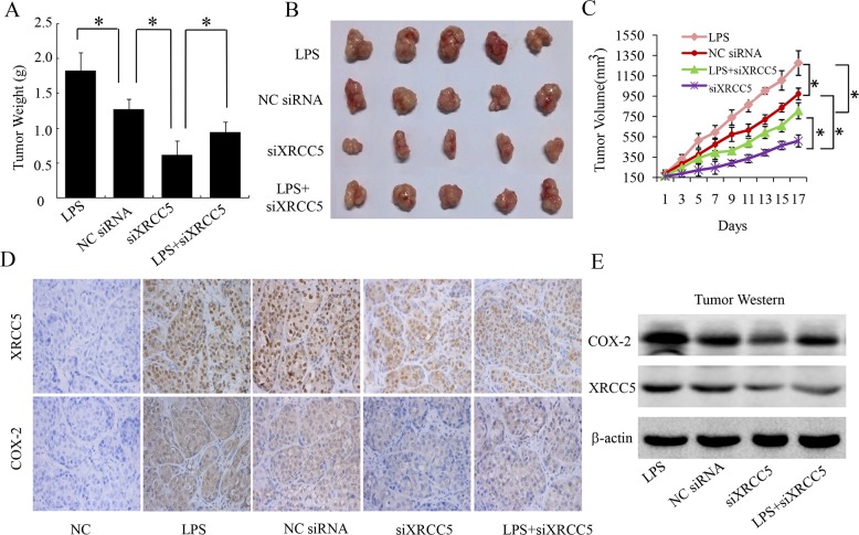
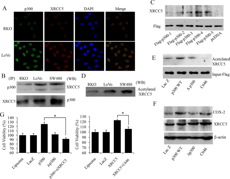
Cyclooxygenase (COX) is the rate-limiting enzyme in prostaglandins (PGs) biosynthesis. Previous studies indicate that COX-2, one of the isoforms of COX, is highly expressed in colon cancers and plays a key role in colon cancer carcinogenesis. Thus, searching for novel transcription factors regulating COX-2 expression will facilitate drug development for colon cancer. In this study, we identified XRCC5 as a binding protein of the COX-2 gene promoter in colon cancer cells with streptavidin-agarose pulldown assay and mass spectrometry analysis, and found that XRCC5 promoted colon cancer growth through modulation of COX-2 signaling. Knockdown of XRCC5 by siRNAs inhibited the growth of colon cancer cells in vitro and of tumor xenografts in a mouse model in vivo by suppressing COX-2 promoter activity and COX-2 protein expression. Conversely, overexpression of XRCC5 promoted the growth of colon cancer cells by activating COX-2 promoter and increasing COX-2 protein expression. Moreover, the role of p300 (a transcription co-activator) in acetylating XRCC5 to co-regulate COX-2 expression was also evaluated. Immunofluorescence assay and confocal microscopy showed that XRCC5 and p300 proteins were co-located in the nucleus of colon cancer cells. Co-immunoprecipitation assay also proved the interaction between XRCC5 and p300 in nuclear proteins of colon cancer cells. Cell viability assay indicated that the overexpression of wild-type p300, but not its histone acetyltransferase (HAT) domain deletion mutant, increased XRCC5 acetylation, thereby up-regulated COX-2 expression and promoted the growth of colon cancer cells. In contrast, suppression of p300 by a p300 HAT-specific inhibitor (C646) inhibited colon cancer cell growth by suppressing COX-2 expression. Taken together, our results demonstrated that XRCC5 promoted colon cancer growth by cooperating with p300 to regulate COX-2 expression, and suggested that the XRCC5/p300/COX-2 signaling pathway was a potential target in the treatment of colon cancers.


Conflict of interest statement

Competing Interests: We have the following interests: Wenlin Huang and Wuguo Deng are employed by Guangzhou Double Bioproduct Inc. There are no patents, products in development or marketed products to declare. This does not alter our adherence to all the PLOS ONE policies on sharing data and materials.

Figures

Fig 1
Fig 1. XRCC5 binding at the promoter region of COX-2 in colon cancer cells.
(A) Streptavidin-biotin pulldown assay and SDS-PAGE with silver stain. A protein band with a molecular weight of 90-100kDa is indicated with an arrow in the figure. (B) Western blot with a specific antibody against XRCC5 in the pulled down proteins of RKO, LoVo, DLD-1 and SW480 cells. (C) Correlation between XRCC5 and COX-2 expression in protein level evaluated with Western blot in RKO, LoVo, DLD-1 and SW480 cells. (D) Correlation between XRCC5 and COX-2 expression in mRNA level evaluated with RT-PCR in RKO, LoVo, DLD-1 and SW480 cells.
Fig 2
Fig 2. XRCC5 regulating COX-2 promoter activation and protein expression in colon cancer cells.
(A) Left: Western blot of XRCC5 and COX-2 in LoVo cells. Right: Western blot of XRCC5 and COX-2 in RKO cells. (B) Luciferase reporter assay of the activity of COX-2 promoter in LoVo cells. Protein weight is used to adjust relative luciferase activity (RLU), and cells treated with BPS negative control are also used for data alignment. Data in the figure are presented as the meanmoter iP<0.05). (C) Luciferase reporter assay of the activity of COX-2 promoter in RKO cells. Protein weight is used to adjust relative luciferase activity (RLU). Data are presented as the meanve proP<0.05).(D) MTS cell viability assay of RKO cells. Cells treated with LacZ is used for data alignment. Data are presented as the mean±SD. (*P<0.05). Si1, Si2 and Si3 represent three sequences of siRNAs of XRCC5, Sictr represents negative control siRNA of XRCC5, LacZ represents negative vector control, LPS represents lipopolysaccharides, PBS represents PBS negative control, XRCC5 represents overexpression of XRCC5, and CB represents celecoxib.
Fig 3
Fig 3. XRCC5 regulating colon cancer cell proliferation in vitro.
(A) MTS cell viability assay of LoVo cells. Cells treated with BPS negative control are used for data alignment. Data are presented as the meaen.D. (*P<0.05). (B) MTS cell viability assay of RKO cells. Cells treated with PBS negative control are used for data alignment. Data are presented as the meannt.D. (*P<0.05). (C) Morphology observation of LoVo cells. (D) Colony formation assay of LoVo cells. Si1, Si2 and Si3 represent three sequences of siRNAs of XRCC5, Sictr represents negative control siRNA of XRCC5, PBS represents PBS negative control, XRCC5 represents overexpression of XRCC5, and LacZ represents negative control vector.
Fig 4
Fig 4. Knockdown of XRCC5 inhibiting tumor growth by down-regulating COX-2 expression in a colon cancer mouse model.
(A) Tumor weight at the time of sacrifice of mice in each group. Data are presented as the meane.D. (*P<0.05). (B) Morphology images of each tumor xenograft resected from nude mice seventeen days after first treatment in each group. (C) Tumor growth curves. Tumor volumes at each time point are presented as the mean).D. Tumor growth curves are depicted with tumor volume at each time point. (*P<0.05) (D) Tumor tissue immunohistochemistry of XRCC5 and COX-2. (E) Western blot of XRCC5 and COX-2 with nuclear proteins exacted from tumor tissues. LPS represents lipopolysaccharides, NCsiRNA represents negative control siRNA, and siXRCC5 represents knockdown of XRCC5 with siRNAs.
Fig 5
Fig 5. XRCC5 interacting with p300 to co-regulate COX-2 expression in colon cancer cells.
(A) Immunofluorescence and confocal microscopy of XRCC5 and p300 in RKO and LoVo cells. XRCC5 is stained by TRITC-conjugated secondary antibodies (red), p300 is stained by FITC-conjugated secondary antibodies (green), and nuclei are stained with DAPI (blue). (B) Co-immunoprecipitation assay of p300 and XRCC5 in RKO, LoVo and SW480 cells.Left: Immunoprecipitation assay (IP) of p300 and XRCC5. Right: Western blot (WB) of XRCC5 and p300. (C) Bottom: The design of the flag-tagged plasmids with different domains of p300. Left: The interaction between XRCC5 and the different domains of p300 detected by immunoprecipitation assay and Western blot. (D)Western blot of XRCC5 with the nuclear extractsimmunoprecipitated by an anti-acetylation antibody in RKO, LoVo and SW480 cells. (E) Western blot of XRCC5 with the nuclear extracts immunoprecipitated by an anti-acetylation antibody in LoVo cells. (F) Western blot of XRCC5 and COX-2 in LoVo cells. (G) MTS cell viability assay in LoVo cells (Left) and RKO cells (Right). Cells treated with liposome negative control is used for data alignment. Data are presented as the meanen.D. (*P<0.05). lacZ represents negative control vector, p300WT represents wild type p300 overexpression, Δp300 represents histone acetyltransferase (HAT) domain deletion mutant p300, C646 represents p300 HAT inhibitor C646, and siXRCC5 represents knockdown of XRCC5 with siRNAs.

References

    1. Brenner H, Kloor M, Pox CP. Colorectal cancer. Lancet. 2014; 383(9927):1490–1502. doi: 10.1016/S0140-6736(13)61649-9 - DOI - PubMed
    1. Siegel RL, Miller KD, Jemal A. Cancer Statistics, 2017. CA Cancer J Clin. 2017; 67(1):7–30. doi: 10.3322/caac.21387 - DOI - PubMed
    1. Prados J, Melguizo C, Ortiz R, Perazzoli G, Cabeza L, Alvarez PJ, et al. Colon cancer therapy: recent developments in nanomedicine to improve the efficacy of conventional chemotherapeutic drugs. Anticancer Agents Med Chem. 2013; 13(8):1204–1216. - PubMed
    1. Ciombor KK, Wu C, Goldberg RM. Recent Therapeutic Advances in the Treatment of Colorectal Cancer. Annual Review of Medicine. 2015; 66:83–95. doi: 10.1146/annurev-med-051513-102539 - DOI - PubMed
    1. Rouzer CA, Marnett LJ. Structural and functional differences between cyclooxygenases: fatty acid oxygenases with a critical role in cell signaling. Biochem Biophys Res Commun. 2005;338(1):34–44. doi: 10.1016/j.bbrc.2005.07.198 - DOI - PubMed