Review
. 2018 Jan;144(1):7-34.
doi: 10.1111/jnc.14242. Epub 2017 Nov 20.

Nutrient-driven O-GlcNAc in proteostasis and neurodegeneration

Affiliations
Review

Nutrient-driven O-GlcNAc in proteostasis and neurodegeneration

Ilhan Akan et al. J Neurochem. 2018 Jan.

Abstract

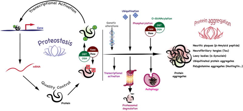
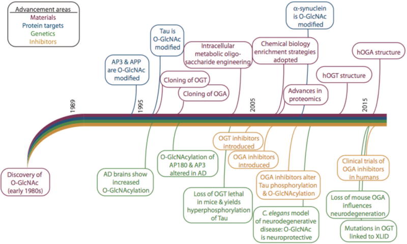
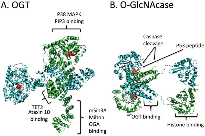
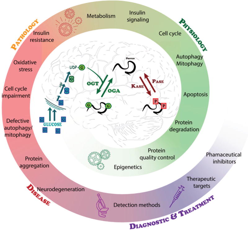
Proteostasis is essential in the mammalian brain where post-mitotic cells must function for decades to maintain synaptic contacts and memory. The brain is dependent on glucose and other metabolites for proper function and is spared from metabolic deficits even during starvation. In this review, we outline how the nutrient-sensitive nucleocytoplasmic post-translational modification O-linked N-acetylglucosamine (O-GlcNAc) regulates protein homeostasis. The O-GlcNAc modification is highly abundant in the mammalian brain and has been linked to proteopathies, including neurodegenerative diseases such as Alzheimer's, Parkinson's, and Huntington's. C. elegans, Drosophila, and mouse models harboring O-GlcNAc transferase- and O-GlcNAcase-knockout alleles have helped define the role O-GlcNAc plays in development as well as age-associated neurodegenerative disease. These enzymes add and remove the single monosaccharide from protein serine and threonine residues, respectively. Blocking O-GlcNAc cycling is detrimental to mammalian brain development and interferes with neurogenesis, neural migration, and proteostasis. Findings in C. elegans and Drosophila model systems indicate that the dynamic turnover of O-GlcNAc is critical for maintaining levels of key transcriptional regulators responsible for neurodevelopment cell fate decisions. In addition, pathways of autophagy and proteasomal degradation depend on a transcriptional network that is also reliant on O-GlcNAc cycling. Like the quality control system in the endoplasmic reticulum which uses a 'mannose timer' to monitor protein folding, we propose that cytoplasmic proteostasis relies on an 'O-GlcNAc timer' to help regulate the lifetime and fate of nuclear and cytoplasmic proteins. O-GlcNAc-dependent developmental alterations impact metabolism and growth of the developing mouse embryo and persist into adulthood. Brain-selective knockout mouse models will be an important tool for understanding the role of O-GlcNAc in the physiology of the brain and its susceptibility to neurodegenerative injury.

Keywords: Alzheimer's; Glucose; Neurodegeneration; O-GlcNAc; O-linked N-acetylglucosamine; Therapeutics.


Conflict of interest statement

The authors have no conflicts to declare

Figures

Figure 1
Figure 1. The potential roles of O-GlcNAcylation in maintaining the cellular proteostasis network and relationship to protein aggregates in neurodegeneration
O-GlcNAcylation has been shown to be a key regulator of transcription, translation, protein folding and degradation that may be regulated in in neurodegenerative disease. The protein aggregates that are associated with human disease are listed at the right. O-GlcNAcylation can influence aggregation by modulating processes including transcription, protein folding, the cellular quality control, and stress pathways. Acting in consort with other post-translational modifications, O-GlcNAc may influence signaling pathways and the regulation of protein degradation including proteasomal degradation, autophagy, and the cellular stress response.
Figure 2
Figure 2. A timeline summarizing the major advancements in understanding O-GlcNAc as a modulator of neurodegeneration
The timeline was derived from publication dates of these references and references therein: (Torres and Hart, 1984; Holt and Hart, 1986; Davis and Blobel, 1987; Hanover et al., 1987; Holt et al., 1987; Park et al., 1987; Snow et al., 1987; D’Onofrio et al., 1988; Caillet-Boudin et al., 1989; Datta et al., 1989; Inaba and Maede, 1989; Haltiwanger et al., 1990; Starr and Hanover, 1990; Starr et al., 1990; Kearse and Hart, 1991a; Kearse and Hart, 1991b; Lüthi et al., 1991; Chou et al., 1992; Hagmann et al., 1992; Haltiwanger et al., 1992; Reason et al., 1992; Roquemore et al., 1992; Dong et al., 1993; Elliot et al., 1993; Kelly et al., 1993; Murphy et al., 1994; Bailer et al., 1995; Chou et al., 1995; Griffith and Schmitz, 1995; Griffith et al., 1995; Dong et al., 1996; Roquemore et al., 1996; Kreppel et al., 1997; Lubas et al., 1997; Roos et al., 1997; Haltiwanger et al., 1998; Yao and Coleman, 1998a; Yao and Coleman, 1998b; Akimoto et al., 1999; Griffith and Schmitz, 1999; Hanover et al., 1999; Akimoto et al., 2000; Lubas and Hanover, 2000; Ross et al., 2000; Cole and Hart, 2001; Gao et al., 2001; Hanover, 2001; Rex-Mathes et al., 2001; Zachara et al., 2001; McClain et al., 2002; Vosseller et al., 2002; Wells et al., 2002; Zachara et al., 2002; Akimoto et al., 2003; Gao et al., 2003; Hanover et al., 2003; Iyer et al., 2003; Lefebvre et al., 2003a; Lefebvre et al., 2003b; Love et al., 2003; Marshall et al., 2003; Whelan and Hart, 2003; Zhang et al., 2003; Cieniewski-Bernard et al., 2004; Jínek et al., 2004; Khidekel et al., 2004; Liu et al., 2004a; Robertson et al., 2004; Brickley et al., 2005; Gross et al., 2005; Hanover et al., 2005; Lefebvre et al., 2005; Dorfmueller et al., 2006; Forsythe et al., 2006; März et al., 2006; Nandi et al., 2006; Andrali et al., 2007; Deng et al., 2008; Rexach et al., 2008; Bleckmann et al., 2009; Gambetta et al., 2009a; Lazarus et al., 2009; Liu et al., 2009; Sinclair et al., 2009; Yanagisawa and Yu, 2009; Yuzwa and Vocadlo, 2009; Di Domenico et al., 2010; Rexach et al., 2010; Srikanth et al., 2010; Kim, 2011; Lazarus et al., 2011; Smet-Nocca et al., 2011; Yuzwa et al., 2011; Nakamura et al., 2012; Wang et al., 2012; Yu et al., 2012; Yuzwa et al., 2012; Cameron et al., 2013; Diwu et al., 2013a; Diwu et al., 2013b; Hanover and Wang, 2013; Wang and Hanover, 2013; Graham et al., 2014; Skorobogatko et al., 2014; Taylor et al., 2014; Cha et al., 2015; Mao et al., 2015; Marotta et al., 2015; Akan et al., 2016; Gatta et al., 2016; Gong et al., 2016; Lagerlöf et al., 2016; Lagerlöf et al., 2017; Lewis et al., 2017; Olivier-Van Stichelen et al., 2017)
Figure 3
Figure 3. The structures of OGT, OGA, and the sites of interaction with some of the binding partners of the enzymes of O-GlcNAc cycling
The crystal structures pictured are derived from crystal structures 1W3B and 3PE4 (OGT) (Jínek et al., 2004; Lazarus et al., 2011) and 5UN8 and 3ZJ0 (OGA) (Rao et al., 2013; Li et al., 2017). (A) Structure and interaction partners of dimer ncOGT. OGT has many binding partners that are crucial for cellular processes mediating neuronal homeostasis. The substrate UDP-GlcNAc bound to the active site is shown in red. The positions in the structure corresponding the interaction domains between OGT and these molecules are indicated in brackets B) Structure and binding partners of the long isoform of O-GlcNAcase. The O-GlcNAcylated p53 peptide binds in the active site is highlighted in red. The sites of caspase 3 cleavage are indicated by the arrows. This form of OGA has been shown to interact with OGT and histones. For discussion of these interactions, see associated portions of the text.
Figure 4
Figure 4. A summary of the role of O-GlcNAc in regulating essential brain functions
UDP-GlcNAc, the end product of the nutrient-sensitive HBP, is dynamically used by OGT to catalyze the addition of O-GlcNAc to substrate proteins. This cyclic modification is coordinately regulated with other PTMs such as phosphorylation to regulate the required intricacies of cellular processes. Deregulation of PTMs including O-GlcNAc leads to several pathologies that are associated with neurodegeneration. Methods for detecting O-GlcNAc and understanding its role in these pathologies continue to improve providing a strong foundation for considering targeted therapeutics based on individual or global protein O-GlcNAcylation and the activities of OGT and OGA.

References

    1. Abramowitz LK, Olivier-Van Stichelen S, Hanover JA. Chromosome imbalance as a driver of sex disparity in disease. J Genomics. 2014;2:77–88. - PMC - PubMed
    1. Akan I, Love DC, Harwood KR, Bond MR, Hanover JA. Drosophila O-GlcNAcase Deletion Globally Perturbs Chromatin O-GlcNAcylation. J Biol Chem. 2016;291:9906–9919. - PMC - PubMed
    1. Akimoto Y, Comer FI, Cole RN, Kudo A, Kawakami H, Hirano H, Hart GW. Localization of the O-GlcNAc transferase and O-GlcNAc-modified proteins in rat cerebellar cortex. Brain Res. 2003;966:194–205. - PubMed
    1. Akimoto Y, Kreppel LK, Hirano H, Hart GW. Localization of the O-linked N-acetylglucosamine transferase in rat pancreas. Diabetes. 1999;48:2407–2413. - PubMed
    1. Akimoto Y, Kreppel LK, Hirano H, Hart GW. Increased O-GlcNAc transferase in pancreas of rats with streptozotocin-induced diabetes. Diabetologia. 2000;43:1239–1247. - PubMed

Publication types

MeSH terms