. 2017 May 13;8(41):69508-69519.
doi: 10.18632/oncotarget.17843. eCollection 2017 Sep 19.

Characterization of a novel androgen receptor (AR) coregulator RIPK1 and related chemicals that suppress AR-mediated prostate cancer growth via peptide and chemical screening

Affiliations

Characterization of a novel androgen receptor (AR) coregulator RIPK1 and related chemicals that suppress AR-mediated prostate cancer growth via peptide and chemical screening

Cheng-Lung Hsu et al. Oncotarget. .

Abstract

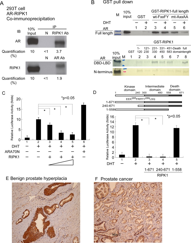
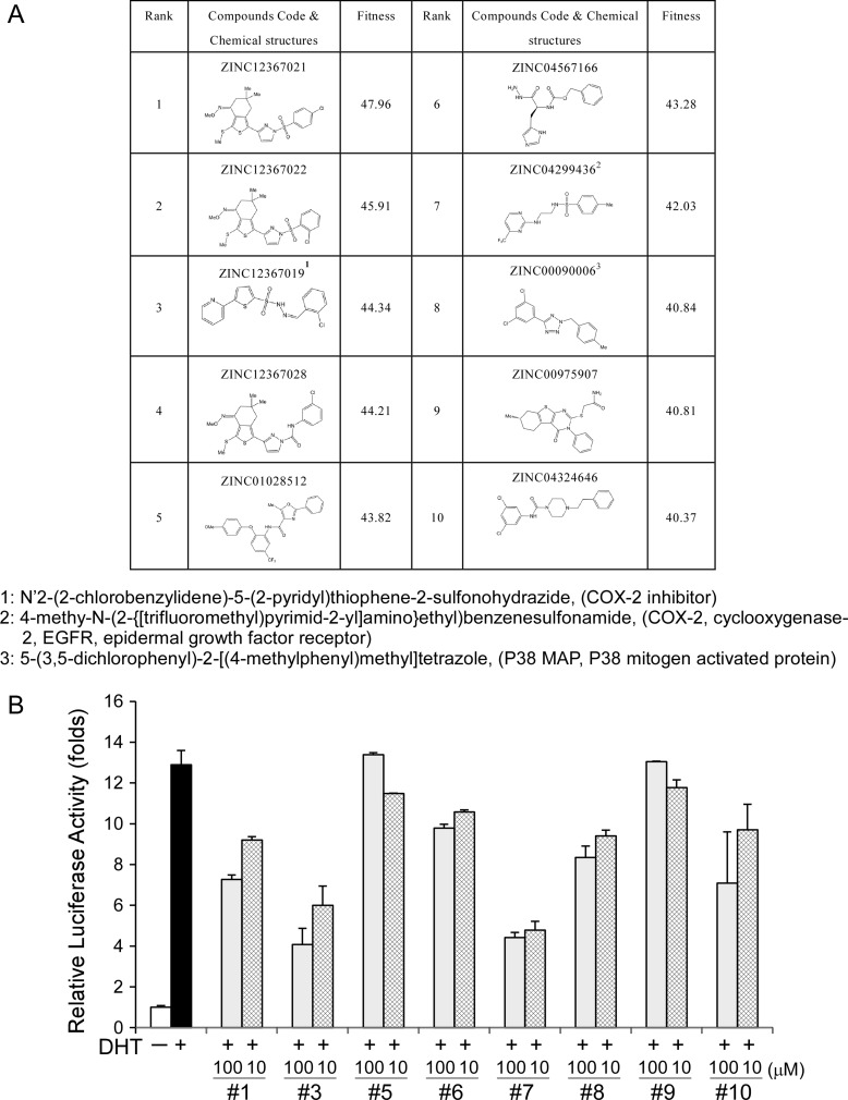
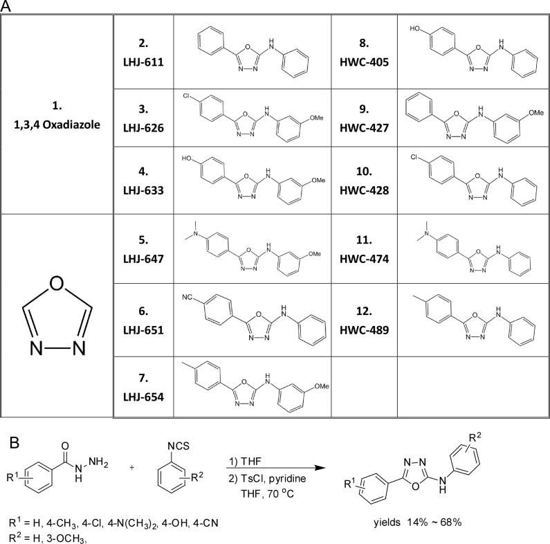
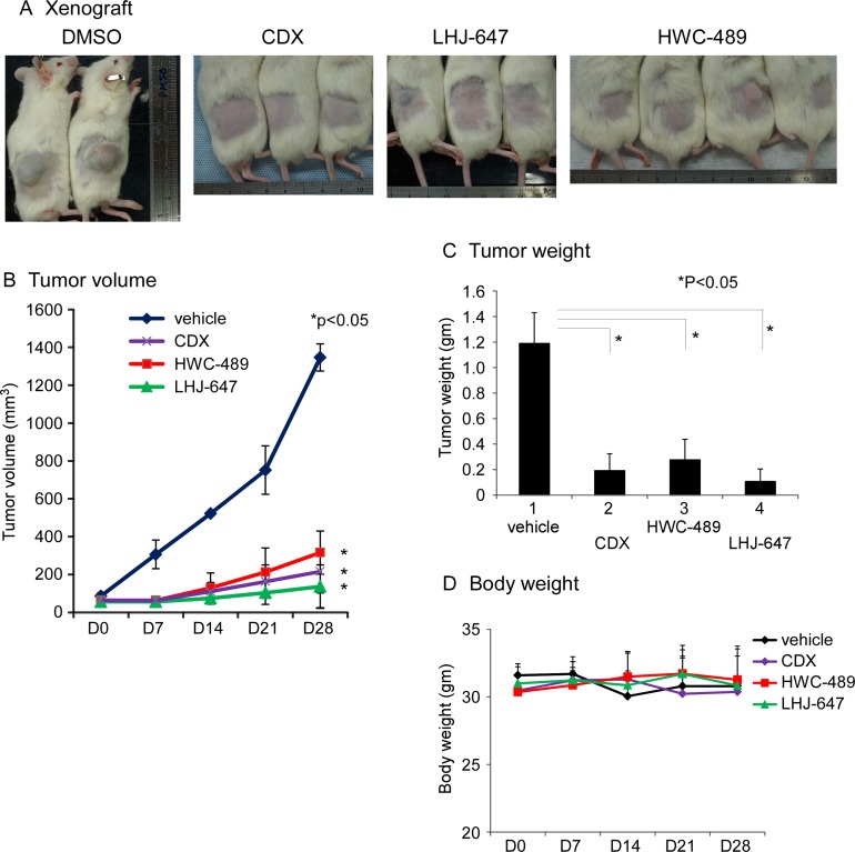
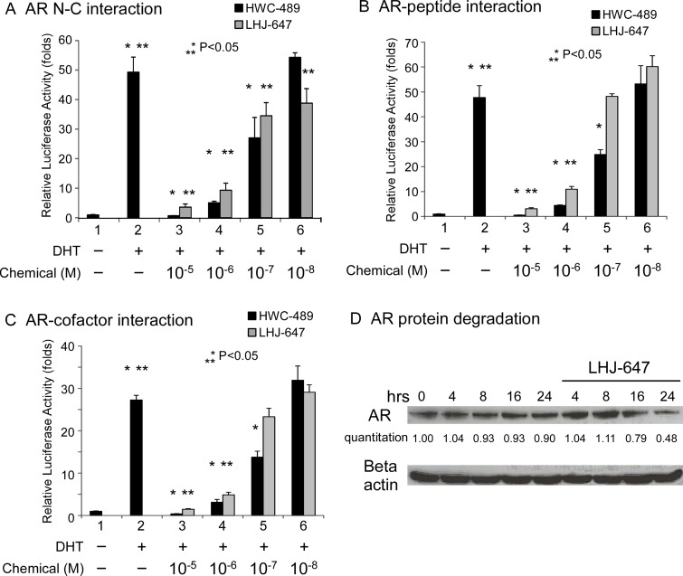
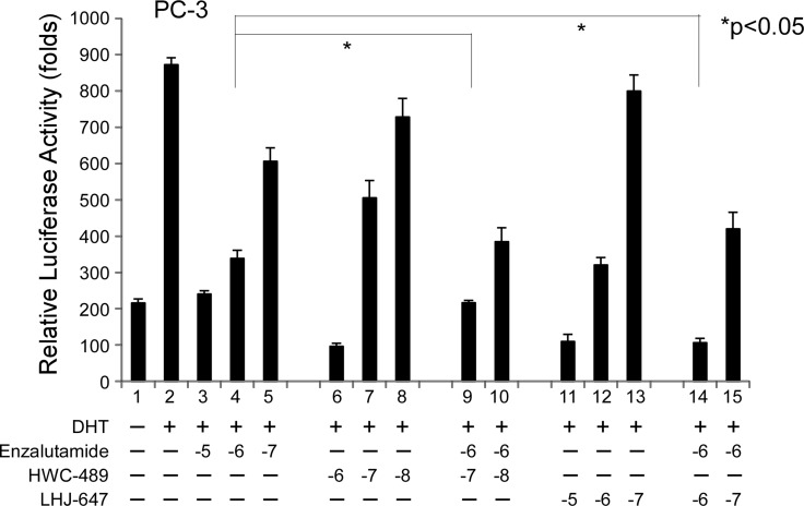
Using bicalutamide-androgen receptor (AR) DNA binding domain-ligand binding domain as bait, we observed enrichment of FxxFY motif-containing peptides. Protein database searches revealed the presence of receptor-interacting protein kinase 1 (RIPK1) harboring one FxxFY motif. RIPK1 interacted directly with AR and suppressed AR transactivation in a dose-dependent manner. Domain mapping experiments showed that the FxxFY motif in RIPK1 is critical for interactions with AR and the death domain of RIPK1 plays a crucial role in its inhibitory effect on transactivation. In terms of tissue expression, RIPK1 levels were markedly higher in benign prostate hyperplasia and non-cancerous tissue regions relative to the tumor area. With the aid of computer modeling for screening of chemicals targeting activation function 2 (AF-2) of AR, we identified oxadiazole derivatives as good candidates and subsequently generated a small library of these compounds. A number of candidates could effectively suppress AR transactivation and AR-related functions in vitro and in vivo with tolerable toxicity via inhibiting AR-peptide, AR-coregulator and AR N-C interactions. Combination of these chemicals with antiandrogen had an additive suppressive effect on AR transcriptional activity. Our collective findings may pave the way in creating new strategies for the development and design of anti-AR drugs.

Keywords: FxxLF; RIPK1; androgen receptor; oxadiazole; prostate cancer.


Conflict of interest statement

CONFLICTS OF INTEREST The authors declare no conflicts of interest.

Figures

Figure 1
Figure 1. Associations between RIPK1 and AR
(A) Co-IP of Flag-RIPK1 with Flag-AR in the 293T cell line. Extracts of 293T cells overexpressing 3xFlag-RIPK1 and 3xFlag-AR were treated with 1 mM DHT. IP was performed using anti-AR (C19) or anti-RIPK1 antibody or normal rabbit serum (negative control), followed by immunoblotting (IB) with antibodies against AR or RIPK1. (B) RIPK1 interacts with full-length AR and the N- and C-terminal regions of AR in GST pull-down assays. Mutation of the FxxFY motif to AxxAA in RIPK1 reduced interactions with AR. RIPK1 suppressed AR transactivation. (C) Transfection of PC-3 prostate cancer cells with AR and RIPK1. PC-3 cells in 24-well plates were co-transfected with 300 ng MMTV-LUC reporter plasmid and 0.5 ng SV40-Renilla luciferase plasmid, together with 100 ng pCMV-Flag-AR and 100, 300 or 500 ng p3xFLAG- RIPK1. The total plasmid DNA content was made up to 1 µg with pCMV. After 16 h, ethanol or 10 nM DHT was added and cells incubated for an additional 16 h. DHT was used as the AR ligand while ARA70N served as the positive control. Relative LUC activity was determined using the dual luciferase system. (D) RIPK1 functional domain mapping in relation to AR transactivation. PC-3 cells were transfected with pCMV-Flag-AR and RIPK1 expression plasmid, P3xFlag-RIPK1 full-length, P3xFlag-RIPK1-(240-671) or P3xflag-RIPK1-(1-558) plasmid, and cultured overnight. Ethanol or 10 nM DHT was added and cells incubated for an additional 16 h. Relative LUC activity was determined using the dual luciferase system. (E) RIPK1 is expressed in benign prostatic hyperplasia tissue, displaying strong positivity (3+, > 90%) in the gland area but weak (1+, 50%) staining in the background. (F) Human prostate cancer tissues were immunostained for RIPK1. “T” indicates the tumor area (right side) and “Non-T” the non-tumor area (left side). RIPK1 expression was weak (1+, 80%) in the cancer area but remained strong (3+, 70%) in the peri-cancer area. The figures are representative of three benign prostatic gland hyperplasia and cancer tissues.
Figure 2
Figure 2. Computer modeling-screened candidate chemicals and their effects on AR transcriptional activity
(A) Candidate chemicals identified from computer modeling and their structures. (B) Examination of the effects of 8 of the top 10 candidates on AR transcriptional activity.
Figure 3
Figure 3. Structures and preparation of oxadiazole and derivatives
(A) Structures of oxadiazole and derivatives (B) Preparation of oxadiazole derivatives. A small molecular compound library of oxadiazole derivatives was prepared as a one-pot synthesis by modification of previously reported procedures [32]. In brief, benzohydrazide was reacted with phenyl isothiocyanate to generate thiosemicarbazide, which was further converted to oxadiazole by the addition of tosyl chloride and pyridine. The yields of the desired products obtained ranged from 14% to 68%.
Figure 4
Figure 4. Oxadiazole derivatives suppress AR-related function in vitro
(A) The candidate chemicals suppressed AR transactivation. The PC-3 prostate cancer cell line was used for experiments. The procedure was similar to that for Figure 1C. Cells were treated with ethanol, DHT or different concentrations of chemicals for 16 h. The candidate chemicals suppressed prostate cancer cell growth in vitro, using CDX as a control, LNCaP (B) CWR22R (C) and PC-3 (D).
Figure 5
Figure 5. Oxadiazole derivatives suppress AR-related function in vivo
(A) The mice with LNCaP cell xenograft after 28 days of treatment with DMSO, CDX, LHJ-647, and HWC-489. (B) Tumor volumes measured during treatment in NOD/SCID mice. HWC-489, LHJ-647, and CDX exerted significant effects, compared to vehicle treatment. (C) Tumor weights measured after sacrificing mice on day 28 of treatment. (D) Mouse body weights of all four groups displayed no significant changes during the treatment period.
Figure 6
Figure 6. Mechanism underlying suppression of AR-related functions by oxadiazole derivatives
(A–C) Transcriptional activity in reporter assays. PC-3 cells in 24-well plates were transfected as indicated below. After incubation for 16 h, cells were treated with ethanol, 10 nM DHT, or 0.01–10 μM HWC-489 or LHJ-647 for an additional 16 h. Luciferase activity in cell lysates was determined and normalized to protein concentrations. Relative luciferase activity was calculated using the luciferase reporter assay system [18]. (A) PC-3 cells were co-transfected with 350 ng pCDNA3-flag-hAR-N (residues 1–506), 350 ng pCDNA3-hAR-C (residues 556–919), and 300 ng MMTV-Luc plasmids. (B) PC-3 cells were co-transfected with 350 ng GAL4-DBD-3-18, 350 ng pCMX-VP16-AR, and 300 ng pG5-Luc plasmids. (C) PC-3 cells were transfected with 350 ng GAL4-DBD-ARA54C, 350 ng VP16-AR, and 300 ng pG5-Luc plasmids. (D) LHJ-647 promotes AR protein degradation.
Figure 7
Figure 7. Addictive effects of oxadiazole derivatives and enzalutamide on AR transcriptional activity
The PC-3 prostate cancer cell line was used for experiments. The procedure used was similar to that for Figure 1C. Cells were treated with ethanol or 10 nM DHT in the absence or presence of different concentrations of candidate chemicals and enzalutamide for 16 h.

References

    1. Miyamoto H, Rahman MM, Chang C. Molecular basis for the antiandrogen withdrawal syndrome. J Cell Biochem. 2004;91:3–12. - PubMed
    1. Chang CS, Kokontis J, Liao ST. Molecular cloning of human and rat complementary DNA encoding androgen receptors. Science. 1988;240:324–26. - PubMed
    1. Lubahn DB, Joseph DR, Sullivan PM, Willard HF, French FS, Wilson EM. Cloning of human androgen receptor complementary DNA and localization to the X chromosome. Science. 1988;240:327–30. - PubMed
    1. Heinlein CA, Chang C. Androgen receptor (AR) coregulators: an overview. Endocr Rev. 2002;23:175–200. - PubMed
    1. Hur E, Pfaff SJ, Payne ES, Grøn H, Buehrer BM, Fletterick RJ. Recognition and accommodation at the androgen receptor coactivator binding interface. PLoS Biol. 2004;2:E274. - PMC - PubMed