. 2017 Jun 9;8(41):69538-69550.
doi: 10.18632/oncotarget.18422. eCollection 2017 Sep 19.

MiR-23a promotes TGF-β1-induced EMT and tumor metastasis in breast cancer cells by directly targeting CDH1 and activating Wnt/β-catenin signaling

Affiliations

MiR-23a promotes TGF-β1-induced EMT and tumor metastasis in breast cancer cells by directly targeting CDH1 and activating Wnt/β-catenin signaling

Fei Ma et al. Oncotarget. .

Abstract

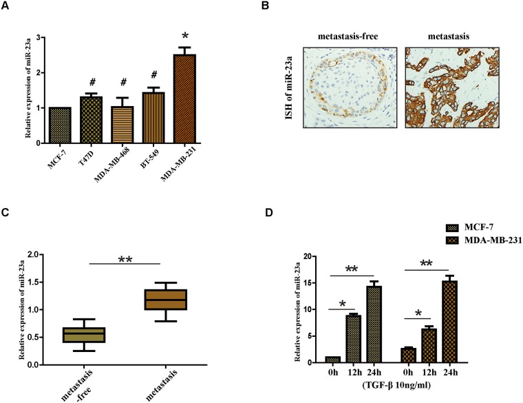
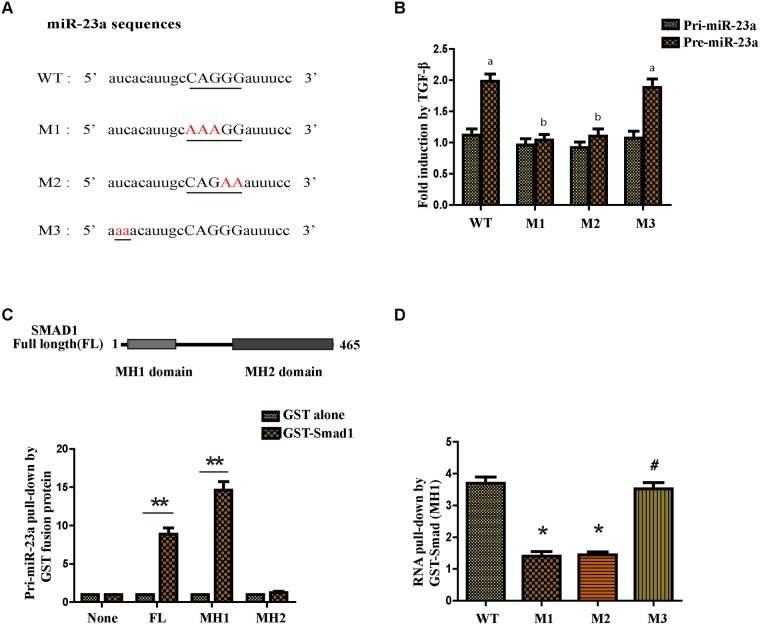
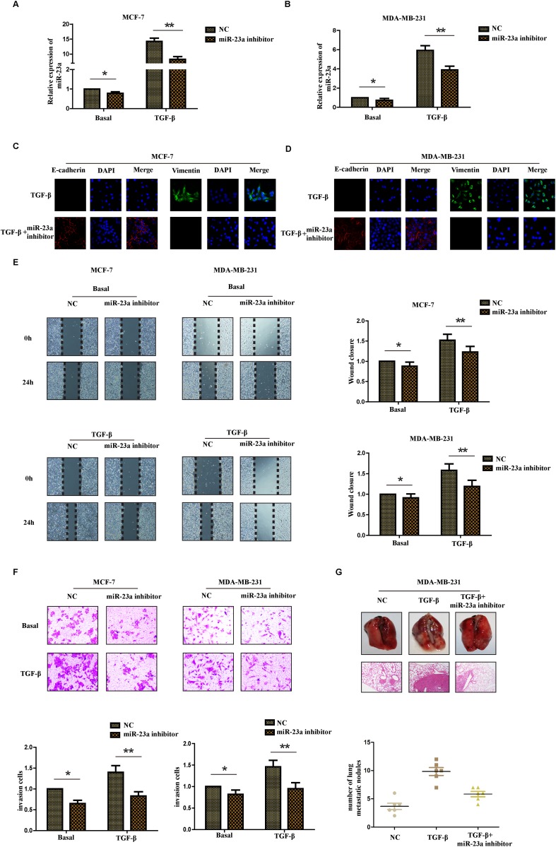
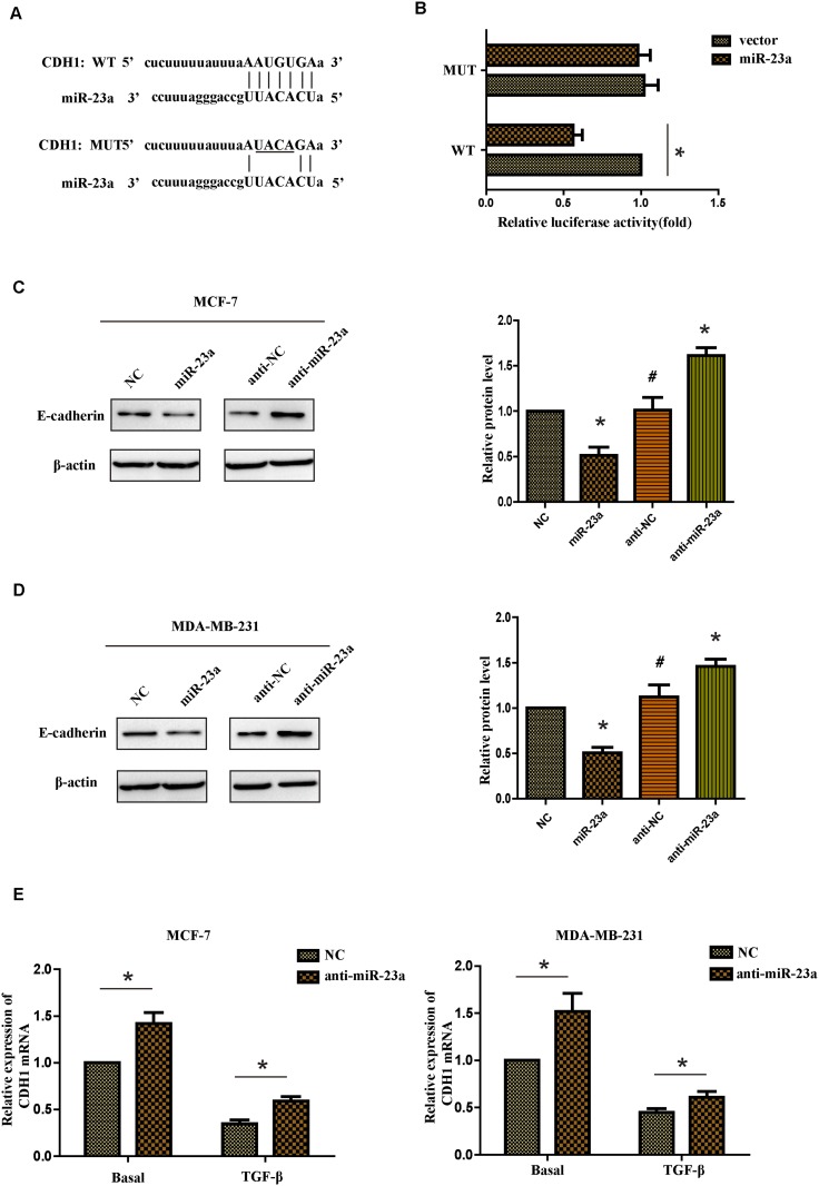
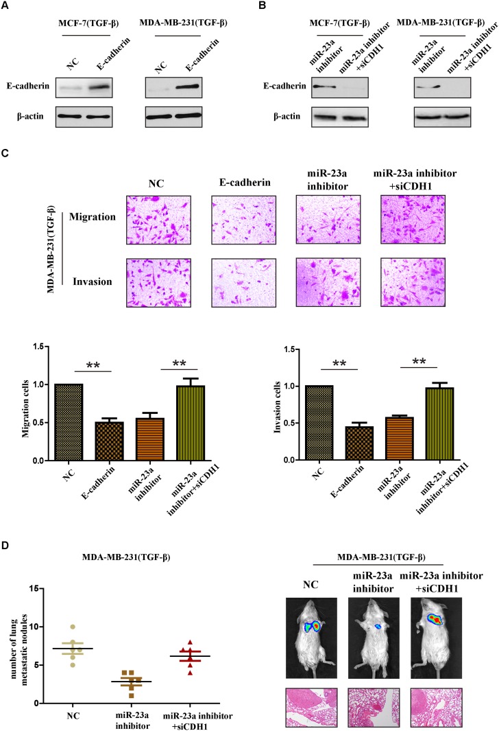
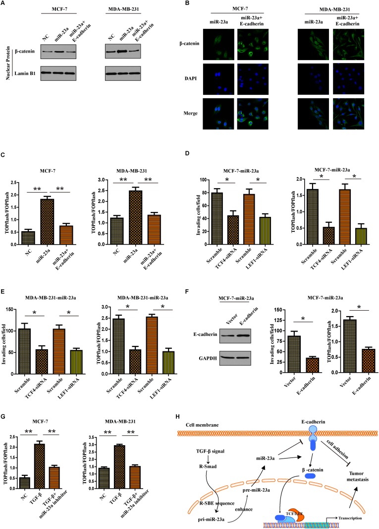
TGF-β1-induced epithelial-mesenchymal transition (EMT) has been proved to be associated with metastasis of breast cancer cells. We attempted to detect a novel mechanism that microRNAs mediated the TGF-β1-induced EMT in the process of breast cancer metastasis. Here we reported that the expression of miR-23a was higher in breast cancer cells with high metastasis ability and patients with lymph node metastasis and the treatment of TGF-β1 significantly upregulated the expression of miR-23a in breast cancer cells. We found that miR-23a was upregulated by TGF-β1 post-transcriptionally and Smads directly bound the RNA Smad binding element (R-SBE) of miR-23a. Functional studies showed that inhibition of miR-23a suppressed the TGF-β1-induced EMT, migration, invasion and metastasis of breast cancer both in vitro and in vivo. In addition, we determined that miR-23a directly targeted and suppressed CDH1, one important gene in EMT phenomenon. Notably, Wnt/β-catenin signaling was activated by the suppression of CDH1 in the miR-23a mediated process of TGF-β1-induced EMT and tumor invasion. These results demonstrate that miR-23a promotes TGF-β1-induced tumor metastasis in breast cancer by targeting CDH1 and activating Wnt/β-catenin signaling. Taken together, our results indicate a novel regulatory mechanism of TGF-β1-induced EMT and suggest that miR-23a might be a potential target in breast cancer therapy.

Keywords: CDH1; R-SBE; TGF-β1; Wnt/β-catenin; miR-23a.


Conflict of interest statement

CONFLICTS OF INTEREST The authors declare no conflicts of interest.

Figures

Figure 1
Figure 1. Expression of miR-23a in cell lines and human tissues and the treatment of TGF-β1 upregulates the expression of miR-23a in MCF-7 and MDA-MB-231 cell lines
(A) Real-time PCR analysis of the expression of miR-23a in different breast cancer cell lines, normalized to GAPDH. (B) Analysis of miR-23a in situ hybridization(ISH) signal in breast cancer tissues. Representative images are shown (400 magnification). (C) Real-time PCR analysis of miR-23a expression in a group of 30 BC patients without lymph node metastasis and matched patients with lymph node metastasis. (D) Real-time PCR analysis of the expression of miR-23a in MCF-7 and MDA-MB-231 cells treated with 10ng/ml TGF-β1 for 12h or 24h. *P<0.05, **P<0.01.
Figure 2
Figure 2. TGF-β1 regulates miR-23a post-transcriptionally and the R-SBE sequence is essential for the association of SMAD MH1 domain and miR-23a
(A) Schematic diagram of pre-miR-23a wild-type and mutant sequences. Underlined characters indicate the R-SBE sequence found in pre-miR-23a and red highlighted characters indicate mutation introduced. (B) MCF-7 cells were transfected with human pri-miR-23a expression constructs, followed by treatment with or without 10ng/ml TGF-β1 for 2h and subjected to real-time PCR analysis using primers to detect exogenous pri-miR-23a or pre-miR-23a, normalized to GAPDH. Fold induction by the TGF-β1 relative to mock treated cells was displayed. Values labeled with different letters differed from one another (P<0.05). (C) The schematic diagram of domains of Smad1 protein (upper panel). In vitro transcribed wild type pri-miR-23a was mixed with indicated recombinant, sepharose bead-immobilized GST-fusion proteins. Associated RNA was eluted, and subjected to real-time PCR analysis to detect pri-miR-23a. The relative amount of pri-miR-23a pulled down with GST-Smad fusion proteins, normalized to the amount pulled down with GST alone is presented (lower panel). (D) In vitro transcribed pri-miR-23a constructs were mixed with recombinant GST-Smad1 (MH1) or GST alone and the relative amount of pri-miR-23a transcripts pulled down with GST-Smad1 (MH1) fusion protein was normalized to the amount pulled down with GST alone. *P<0.05, **P<0.01.
Figure 3
Figure 3. Inhibition of miR-23a suppresses the TGF-β1-induced EMT, the migration and invasion ability of breast cancer cells treated with TGF-β1
Real-time PCR was used to confirm the transfection efficiency of miR-23a in MCF-7 (A) and MDA-MB-231 (B) cells treated with or without TGF-β1. (C and D) Representative IF images indicated that miR-23a had an effect on the expression of EMT genes in indicated cells treated with TGF-β1. (E) Representative micrograph images of wound healing assay of the indicated cells. Wound closures were photographed at 0h and 24h after wounding. (F) Invasion assays of the indicated cells transfected with NC or miR-23a inhibitor. (G) Tumor metastasis analysis. Representative images of lung tissues and micrographs of HE staining of metastatic tumor tissues (upper panel). The number of metastatic lung nodules in each group of nude mice (n= 6 per group, lower panel). *P<0.05, **P<0.01.
Figure 4
Figure 4. CDH1 is a direct target of miR-23a
(A) Luciferase reporter plasmids containing the putative wild-type or mutant miR-23a binding sequence in 3’-UTR of CDH1 mRNA. (B) HEK293T cells were co-transfected with a control vector or miR-23a and a luciferase reporter construct containing the wild-type or mutant CDH1 3’-UTR. The results were normalized, and the luciferase activity of the control was set to 1. (C and D) Effects of miR-23a dysregulation of CDH1 expression were detected by western blot analysis in MCF-7 and MDA-MB-231 cells. (E) Real-time PCR analysis of CDH1 mRNA expression of the NC and anti-miR-23a-transfected cells treated with or without TGF-β1. *P<0.05, **P<0.01.
Figure 5
Figure 5. CDH1 critically contributes to the pro-metastatic function of miR-23a in breast cancer cells with TGF-β1 treatment
(A) Western blot analysis confirmed the transfection of constructs containing CDH1 ORF in cells treated with TGF-β1. (B) Western blot analysis confirmed the transfection of specific siRNA in miR-23a-silenced cells treated with TGF-β1. (C) Reintroduction of E-cadherin in TGF-β1-treated MDA-MB-231 cells abrogated TGF-β1-induced cell invasion. MiR-23a inhibitor reduced the invasion and migration cells treated by TGF-β1 and siCDH1 restored the cell invasion and migration in miR-23a-silenced cells. (D) Tumor metastasis analysis. The number of metastatic lung nodules in each group of nude mice (n= 6 per group, left panel). Representative photos of nude mice injected with indicated cells and micrographs of HE staining of metastatic tumor tissues (right panel). *P<0.05, **P<0.01.
Figure 6
Figure 6. MiR-23a targets CDH1 to hyperactivate Wnt/β-catenin signaling and subsequently mediates the TGF-β1-induced EMT and tumor invasion in breast cancer
(A) Nuclear fraction of indicated cells was analyzed by Western blot. Lamin B1 was used as a loading control. (B) Beta-catenin localization in indicated cells was detected by immunofluorescence staining. (C) Indicated cells were transfected with TOPflash and Renilla pRL-TK plasmids, and subjected to dual –luciferase assays 48h after transfection. Reporter activity was normalized to Renilla luciferase activity. (D and E) Transwell assays were used to detect the quantification of invading cells transfected with indicated siRNAs and luciferase-reported TCF/LEF transcriptional activity in indicated cells were examined. (F) Western blot analysis was used to confirm the transfection of constructs containing CDH1 ORF in miR-23a overexpressed MCF-7 cells (left panel). Quantification of invading cells transfected with indicated construct (middle panel) and luciferase-reported TCF/LEF transcriptional activity in indicated cells (right panel). (G) Luciferase-reported TCF/LEF transcriptional activity in NC, cells treated with TGF-β1 or miR-23a silenced-cells treated with TGF-β1. (H) Schematic representation of a model for the role of miR-23a in the TGF-β-induced tumor metastasis in breast cancer. *P<0.05, **P<0.01.

References

    1. Jemal A, Bray F, Center MM, Ferlay J, Ward E, Forman D. Global cancer statistics. CA Cancer J Clin. 2011;61:69–90. - PubMed
    1. Spano D, Heck C, De Antonellis P, Christofori G, Zollo M. Molecular networks that regulate cancer metastasis. Semin Cancer Biol. 2012;22:234–49. - PubMed
    1. Thiery JP, Acloque H, Huang RY, Nieto MA. Epithelial-mesenchymal transitions in development and disease. Cell. 2009;139:871–90. - PubMed
    1. Thiery JP, Sleeman JP. Complex networks orchestrate epithelial-mesenchymal transitions. Nat Rev Mol Cell Biol. 2006;7:131–42. - PubMed
    1. Ozdamar B, Bose R, Barrios-Rodiles M, Wang HR, Zhang Y, Wrana JL. Regulation of the polarity protein Par6 by TGFbeta receptors controls epithelial cell plasticity. Science. 2005;307:1603–09. - PubMed

LinkOut - more resources