. 2017 Aug 3;8(41):70630-70641.
doi: 10.18632/oncotarget.19906. eCollection 2017 Sep 19.

LncSHRG promotes hepatocellular carcinoma progression by activating HES6

Affiliations

LncSHRG promotes hepatocellular carcinoma progression by activating HES6

Ying-Chen Xu et al. Oncotarget. .

Abstract

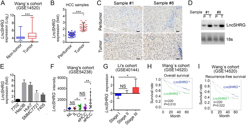
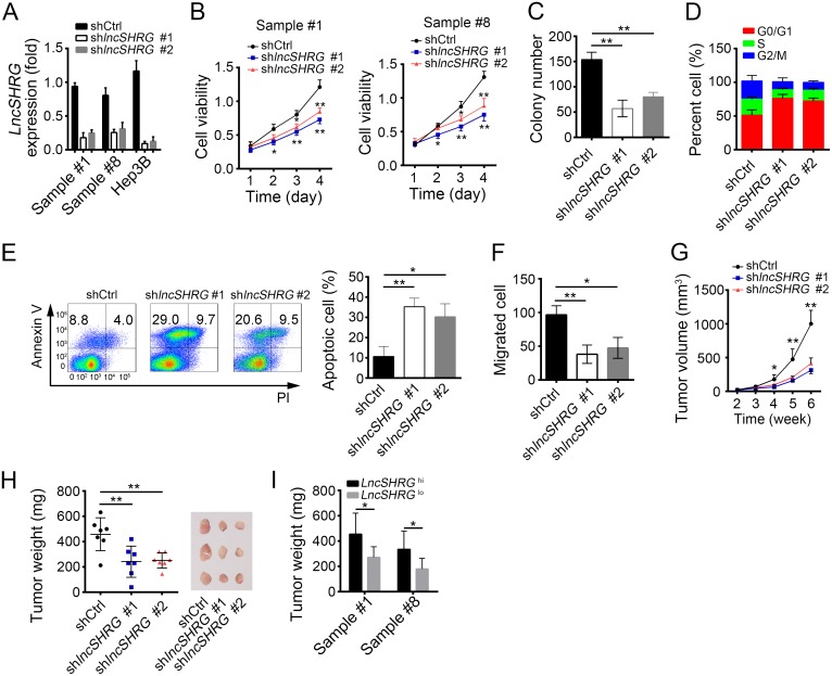
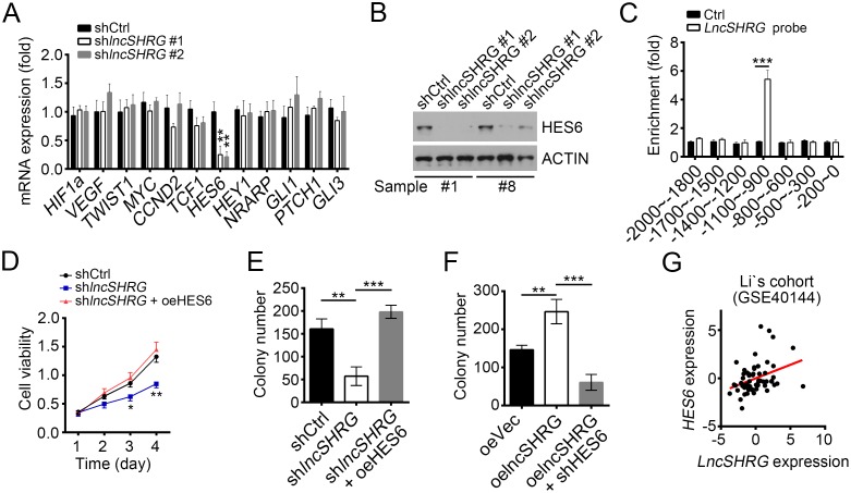
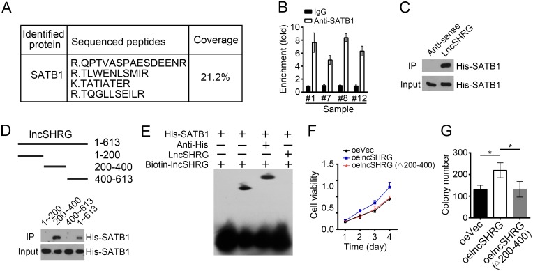
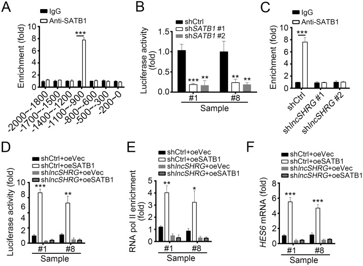
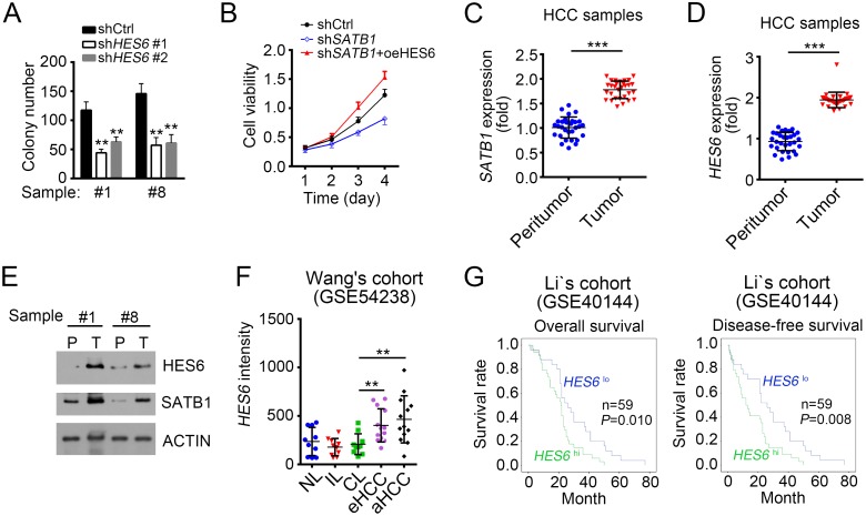
Hepatocellular carcinoma, one of the most common cancers, leads to mass mortality worldwide currently. However, the underlying mechanism of its oncogenesis remains to be elucidated. Here we identified that a long noncoding RNA, lncSHRG, was greatly upregulated in human hepatocellular carcinoma samples. We found that lncSHRG was essential for liver cancer cell proliferation and tumor propagation in mice. In mechanism, lncSHRG recruits SATB1 to bind to HES6 promoter and initiates HES6 expression. HES6, which is highly expressed in hepatocellular carcinoma, promotes tumor cell proliferation. High expression level of HES6 is positively correlated with clinical severity and poor prognosis of people with hepatocellular carcinoma. Altogether, our research provides a new insight on the mechanism of hepatocellular carcinoma progression.

Keywords: HCC; HES6; SATB1; lncSHRG; proliferation.


Conflict of interest statement

CONFLICTS OF INTEREST The authors declare no competing financial interests.

Figures

Figure 1
Figure 1. LncSHRG is highly expressed in HCC
(A) Analysis of lncSHRG expression in peritumor and tumor tissues according to the microarray data in Wang’s cohort (GSE14520). (B) 30 pairs of peritumor and tumor HCC samples were collected. Then LncSHRG expression levels were analyzed in these sample tissues by RT-qPCR. Fold changes were normalized to endogenous ACTB. (C) LncSHRG expression in peritumor and tumor tissues of sample #1 and #8 was checked by RNA hybridization in situ with biotin-labeled lncSHRG probes. Scale bars, 100μm. (D) Total RNAs were extracted from peritumor and HCC samples. LncSHRG and 18S rRNA (loading control) was examined by Northern blot. P: peritumor; T: tumor. (E) Total RNAs were extracted from indicative human HCC cell lines and lncSHRG expression was checked by RT-qPCR. Fold changes were normalized to endogenous ACTB. (F) Higher expression of lncSHRG in HCC samples. LncSHRG expression was analyzed with R language and Bioconductor according to the microarray data in Wang’s cohort (GSE54238). NL: normal livers; IL: chronic inflammatory livers; CL: cirrhotic livers; eHCC: early HCC; aHCC: advanced HCC. (G-I) LncSHRG expression levels were positively correlated with clinical stages and poor prognosis by expression analysis (G) and Kaplan–Meier survival analysis (H and I) according to the microarray data in Li’s cohort (GSE40144) and Wang’s cohort (GSE14520). *p<0.05, **p<0.01 and ***p<0.001 by two-tailed Student’s t test. All data presented are shown as means ± SD collected from three independent experiments.
Figure 2
Figure 2. LncSHRG is required for liver cancer cell proliferation and migration
(A) LncSHRG was knocked down in HCC sample #1 and #8, and Hep3B cells using two independent siRNA sequences by infection of lentivirus containing pSICOR-GFP-shlncSHRG. GFP-positive cells were isolated by FACS and cultured. Knockdown efficiency of lncSHRG was checked by real time qPCR. Cells infected with lentivirus containing pSICOR-GFP-scramble were chosen as control. (B and C) Effect of lncSHRG knockdown on proliferation of HCC samples was measured by MTT assays and colony formation assays. (D) Cell-cycle distribution of HCC sample #1 cells was analyzed after lncSHRG knockdown by FACS. (E) Cell apoptosis was checked in lncSHRG-depleted HCC sample #1 cells by FACS. Cells were stained with Annexin V/PI. (F) Effect of lncSHRG on Hep3B cell migration was analyzed by transwell assays. (G and H) The volumes and weights of tumors were measured at indicated time points. 2×10⁶ lncSHRG-depleted or control Hep3B cells were injected into nude mice. The weights of tumors were measured on week 5 after injection. (I) LncSHRG was important for HCC propagation. Sample cells were classed into 2 groups based on the expression level of lncSHRG. Then 2×10⁶ lncSHRG highly expressed or lowly expressed sample cells were injected into nude mice and tumor weights were measured on week 5. N=6 for each group. *p<0.05 and **p<0.01 by two-tailed Student’s t test. All data presented are shown as means ± SD collected from three independent experiments.
Figure 3
Figure 3. LncSHRG positively regulates liver cancer cell through HES6
(A) LncSHRG knockdown impaired HES6 expression. Indicative gene expression levels in shCtrl or shlncSHRG HCC samples were analyzed by RT-qPCR. (B) HES6 protein levels were downregulated in HCC sample #1 and #8 cells after lncSHRG depletion. (C) LncSHRG bound to HES6 promoter by CHIRP assay. Biotin-labeled lncSHRG probes were designed online and purchased from Invitrogen. HCC sample cells were lysed and genomes were sonicated into 300–500 bp DNA fragments. Then lysates were incubated with lncSHRG probes for enrichment of lncSHRG and DNA fragments. (D and E) Cell proliferation ability was analyzed by MTT assays and colony formation assay.GFP-positive sample #1 cells infected with pSICOR-GFP-shlncSHRG or pSICOR-GFP-scramble were chosen for assays. For HES6 overexpression, sample cells containing pSICOR-GFP-shlncSHRG were transfected with pCDNA3-HES6 plasmid using Lipofectamine 2000 (Invitrogen, USA). (F) HES6 knockdown inhibited colony formation. (G) LncSHRG expression was positively correlated with that of HES6 in HCC samples. *p<0.05, **p<0.01 and ***p<0.001 by two-tailed Student’s t test. All data presented are shown as means ± SD collected from three independent experiments.
Figure 4
Figure 4. LncSHRG associates with SATB1
(A) Biotin-RNA pulldowns were performed using biotin-labeled lncSHRG or anti-sense control. Eluted fractions were resolved by SDS-PAGE, followed by silver staining and mass spectrometry. SATB1 was identified as a potential interactive protein of lncSHRG. (B) HCC sample lysates were incubated with anti-SATB1 at 4°C for 4 h, followed by an RNA immunoprecipitation assay. (C) Interaction of His-SATB1 with biotin-lncSHRG was checked by RNA pulldown assays. (D) lncSHRG (nt200∼400) was essential for the interaction with SATB1 as shown by domain mapping and RNA pulldown assays. (E) Biotin-labeled lncSHRG (nt200∼400) probe was incubated with His-SATB1 protein, followed by EMSA assays. (F) LncSHRG (nt200∼400) contributed to cell proliferation by MTT assays. Full-length lncSHRG but not truncation version (deletion of nt200∼400) promotes cell proliferation in HCC samples. (G) LncSHRG (nt200∼400) promoted colony formation. Full-length lncSHRG but not truncation version (deletion of nt200∼400) promotes colony formation of HCC sample cells. *p<0.05 by two-tailed Student’s t test. All data presented are shown as means ± SD collected from three independent experiments.
Figure 5
Figure 5. LncSHRG is essential for SATB1 binding to HES6 promoter
(A) SATB1 bound to HES6 promoter (-1100bp∼-900bp) as shown by ChIP assays. χ axis stands for the distance from transcription start site (TSS). (B) SATB1 knockdown inhibited HES6 transcription. Luciferase activity assays were performed with SATB1-silenced sample cells. (C) LncSHRG knockdown impaired SATB1 binding to HES6 promoter (-1100∼-900) as shown by ChIP-qPCR assays. WT or lncSHRG-silenced HCC sample cells were lysed and incubated with anti-SATB1 for ChIP assays. (D) LncSHRG was essential for SATB1-regulated HES6 transcription. Luciferase activity assays were performed with WT or lncSHRG-silenced sample cells. (E) SATB1 overexpression promoted enrichment of RNA pol II on HES6 promoter (-1100∼-900) via an lncSHRG-dependent manner. (F) SATB1 promoted HES6 expression relying on the presence of lncSHRG. WT or lncSHRG-silenced sample cells were transfected with SATB1-overexpressing plasmid or empty plasmid control. Then mRNA levels of HES6 were measured by real time qPCR. *p<0.05, **p<0.01 and ***p<0.001 by two-tailed Student’s t test. All data presented are shown as means ± SD collected from three independent experiments.
Figure 6
Figure 6. HES6 promotes proliferation and is positively correlated with poor prognosis
(A) HES6 knockdown inhibited colony formation. HES6 was knocked down for colony formation assays. (B) HES6 overexpression promoted cell proliferation. WT or SATB1-silenced sample cells were transfected with HES6-overexpressing plasmid or control. Then MTT assays were conducted. (C and D) SATB1 and HES6 were expressed higher in tumor than peritumor as measured by RT-qPCR. (E) SATB1 and HES6 expression levels were checked by WB in HCC samples. (F) High expression of HES6 in serious HCC samples as analyzed according to the microarray data in Wang’s cohort (GSE54238). (G) HES6 expression is positively correlated with HCC poor prognosis. Kaplan–Meier survival analyses were performed according to the microarray data in Li’s cohort (GSE40144). *p<0.05, **p<0.01 and ***p<0.001 by two-tailed Student’s t test. All data presented are shown as means ± SD collected from three independent experiments.

References

    1. Bruix J, Gores GJ, Mazzaferro V. Hepatocellular carcinoma: clinical frontiers and perspectives. Gut. 2014;63:844–55. - PMC - PubMed
    1. Wang YY, Qi LN, Zhong JH, Qin HG, Ye JZ, Lu SD, Ma L, Xiang BD, Li LQ, You XM. High expression of AKR1B10 predicts low risk of early tumor recurrence in patients with hepatitis B virus-related hepatocellular carcinoma. Sci Rep. 2017;7:42199. - PMC - PubMed
    1. Correnti M, Raggi C. Stem-like plasticity and heterogeneity of circulating tumor cells: current status and prospect challenges in liver cancer. Oncotarget. 2017;8:7094–115. https://doi.org/10.18632/oncotarget.12569. - DOI - PMC - PubMed
    1. Oikawa T. Cancer stem cells and their cellular origins in primary liver and biliary tract cancers. Hepatology. 2016;64:645–51. - PubMed
    1. Morise Z, Kawabe N, Tomishige H, Nagata H, Kawase J, Arakawa S, Yoshida R, Isetani M. Recent advances in the surgical treatment of hepatocellular carcinoma. World J Gastroenterol. 2014;20:14381–92. - PMC - PubMed

LinkOut - more resources