Review
. 2018 Jan 1;124(1):208-224.
doi: 10.1152/japplphysiol.00747.2017. Epub 2017 Oct 19.

Exercise limitations in heart failure with reduced and preserved ejection fraction

Affiliations
Review

Exercise limitations in heart failure with reduced and preserved ejection fraction

David C Poole et al. J Appl Physiol (1985). .

Abstract

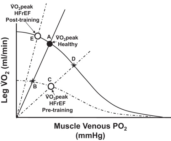
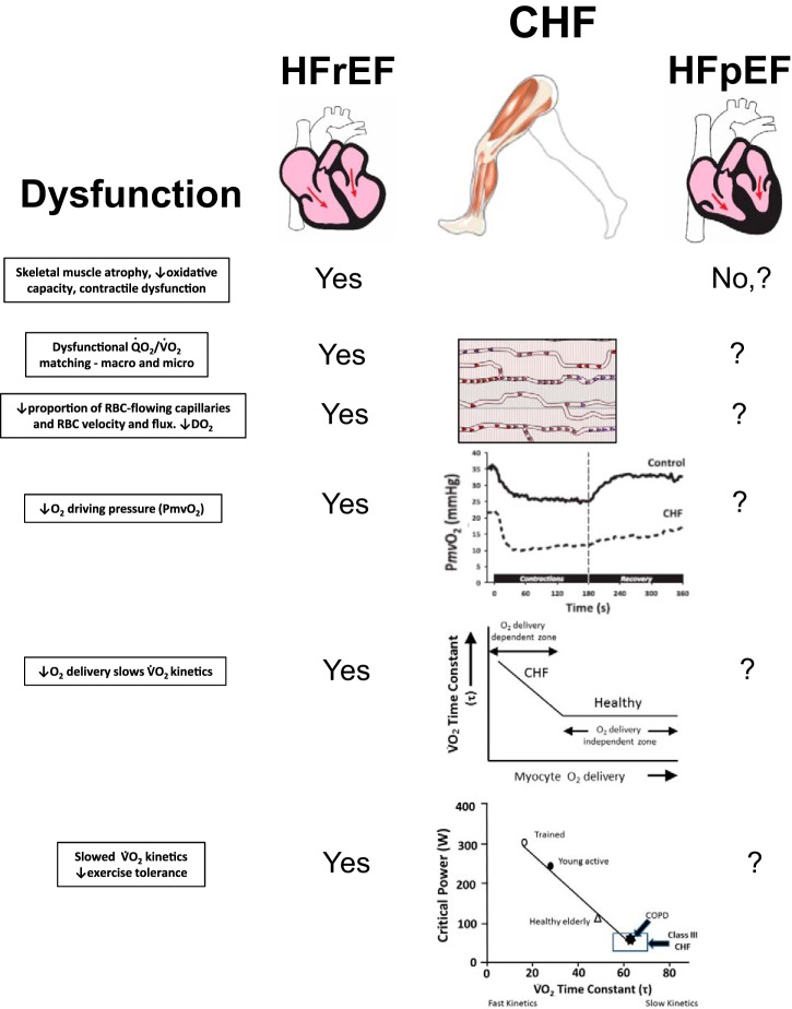
The hallmark symptom of chronic heart failure (HF) is severe exercise intolerance. Impaired perfusive and diffusive O2 transport are two of the major determinants of reduced physical capacity and lowered maximal O2 uptake in patients with HF. It has now become evident that this syndrome manifests at least two different phenotypic variations: heart failure with preserved or reduced ejection fraction (HFpEF and HFrEF, respectively). Unlike HFrEF, however, there is currently limited understanding of HFpEF pathophysiology, leading to a lack of effective pharmacological treatments for this subpopulation. This brief review focuses on the disturbances within the O2 transport pathway resulting in limited exercise capacity in both HFpEF and HFrEF. Evidence from human and animal research reveals HF-induced impairments in both perfusive and diffusive O2 conductances identifying potential targets for clinical intervention. Specifically, utilization of different experimental approaches in humans (e.g., small vs. large muscle mass exercise) and animals (e.g., intravital microscopy and phosphorescence quenching) has provided important clues to elucidating these pathophysiological mechanisms. Adaptations within the skeletal muscle O2 delivery-utilization system following established and emerging therapies (e.g., exercise training and inorganic nitrate supplementation, respectively) are discussed. Resolution of the underlying mechanisms of skeletal muscle dysfunction and exercise intolerance is essential for the development and refinement of the most effective treatments for patients with HF.

Keywords: HFpEF; HFrEF; diffusive O2 transport; exercise intolerance; exercise training; heart failure; perfusive O2 transport; skeletal muscle microcirculation.


Figures

Fig. 1.
Fig. 1.
Oxygen uptake (V̇o2) plotted as a function of venous or microvascular Po2 (Wagner diagram). Top: a schematic illustration of the perfusive [V̇o2 =  O2 (CaCvO2)] and diffusive (V̇o2 = DO2 × Po2) components that interact to determine peak oxygen uptake (V̇o2max). Note that the Fick principle line is not straight because it is directly reflective of the hemoglobin dissociation curve. Therefore, a left-shifted hemoglobin dissociation curve (greater hemoglobin O2 affinity), resulting in a lower venous Po2, will bisect the Fick law line earlier and will reduce V̇o2max and vice versa. The slope of the straight line emanating from the origin is determined by the diffusing capacity (DO2) of the muscle(s). What is often not appreciated within the Wagner diagram is that fractional O2 extraction (CaCvO2) will be determined by the ratio of muscle diffusing capacity (DO2) to blood flow () according to CaCvO2 = V̇o2/O2 =  O2 (1eDO2/βQ˙), where β is the slope of the O2 dissociation curve in the physiological range (105). That the ratio DO2/ determines fractional O2 extraction explains how a high CaCvO2 can be preserved by a very low even in the face of a pathologically reduced DO2 as in HFrEF. Bottom: V̇o2max is plotted for individuals with chronic heart failure (HFrEF) and healthy control subjects. Note that the patients with HF (HFrEF) exhibit attenuated perfusional (Fick principle curve shifted downward) and diffusional (decreased slope of the Fick law line) conductances that contribute to the significant reductions in V̇o2max.
Fig. 2.
Fig. 2.
A schematic representation of the relatively small ratio of cardiac capacity to skeletal muscle recruitment during standard cycle ergometry (top), the much greater ratio during single-leg knee extension (bottom), and the subsequently contrasting physiological responses to these exercise paradigms. Arrows designate relative change from resting.
Fig. 3.
Fig. 3.
Top: schematic illustration of Fick’s law applied to blood-myocyte O2 transport within the skeletal muscle microcirculation. Po2mv constitutes the main driving pressure for transmembrane O2 flux given that intracellular Po2 (Po2intracel) approximates 0 during contractions [approximately 1–3 mmHg; particularly during maximal exercise (101)]. The direct consequence of reduced Po2mv with HFrEF is thus impaired blood-myocyte O2 transport. [Adapted from Hirai et al. (57).] DO2m, muscle O2 diffusing capacity; in, interstitium; p, plasma; RBC, red blood cell; V̇o2; O2 flux. See main text for further details. Bottom: spinotrapezius muscle microvascular Po2 (Po2mv) profiles during the rest-contraction transient in healthy and HF rats with reduced ejection fraction (HFrEF). Note that HFrEF speeds the kinetics of Po2mv fall, thus lowering muscle microvascular oxygenation across the transient (blue area). Time 0 denotes the onset of contractions. Arrow designates the lowering of microvascular Po2 by HFrEF during contractions. [Adapted from Poole et al. (95b).]
Fig. 4.
Fig. 4.
A schematic representation of the perfusive (O2) and diffusive (DO2) components that interact to determine V̇o2peak in patients with HFrEF and healthy controls during both cycle (large muscle mass) and knee-extensor (KE; small muscle mass) exercise. Note, if reductions in V̇o2peak in HFrEF were entirely due to reductions in O2, then the response would follow from point A to B (without any change in the DO2). However, the reductions in V̇o2peak for both cycle and KE exercise in HFrEF were found to follow from point A to C. With this scenario, reductions in DO2 also contributed to the reductions in V̇o2peak in HFrEF as demonstrated by the changes in the slope of the line from B to C. These reductions in DO2 occurred in HFrEF without any demonstrable change in muscle venous Po2 (or muscle venous O2 content) and thus without any changes in arterial-venous O2 content difference.
Fig. 5.
Fig. 5.
Improvement in skeletal muscle diffusional conductance (DO2) in relation to leg V̇o2peak during maximal cycle and knee-extensor (KE) exercise afforded by 8 wk of KE training in patients with HFrEF compared with healthy sedentary controls. Note, the correlation coefficient only represents the relationship between the individual data.
Fig. 6.
Fig. 6.
A schematic illustration of the perfusive and diffusive components that interact to determine leg V̇o2peak in both HF (HFrEF) and control subjects during cycle (large muscle mass) and knee-extensor exercise (KE; small muscle mass) and the subsequent changes as a consequence of KE training. Solid lines represent Fick’s law and principle lines for the healthy sedentary controls (intersecting at A; V̇o2peak Healthy), whereas the dotted lines represent the patients with HFrEF. Following training, both groups share the solid Fick principle line. In both exercise modalities, before training, the patients with HFrEF exhibited attenuated perfusive and diffusive oxygen transport as evidenced by their V̇o2peak being defined by the intercept of lower Fick’s law and principle lines (C) with B indicating the less severe reduction in V̇o2peak had the patients only revealed a reduction in perfusive O2 transport. KE training corrected both of these deficits, without increasing cardiac output, restoring both skeletal muscle perfusive and diffusive O2 transport and therefore allowing V̇o2peak to equal or exceed (KE) that of the healthy sedentary controls (E). D represents the consequence of exercise training if the increase in V̇o2peak had only been driven by an increase in perfusive O2 transport (C to D).
Fig. 7.
Fig. 7.
Top: spinotrapezius muscle microvascular Po2 (Po2mv) profiles during the rest-contractions transient in sedentary and endurance exercise-trained HF rats with reduced ejection fraction (HFrEF). Time 0 denotes the onset of contractions. SE bars are omitted for clarity. The inset displays the overall dynamics of Po2mv fall during contractions (mean response time; MRT) in sedentary (S) and exercise-trained (T) HFrEF rats. Note that exercise training slows Po2mv kinetics (↑MRT), thus increasing muscle microvascular oxygenation across the transient (red area). Gray arrow demonstrates training effect. Bottom: changes in muscle microvascular oxygenation (ΔPo2area) with SNP and l-NAME in sedentary and exercise-trained HFrEF rats. ΔPo2area is calculated as the difference in the area under the Po2mv curve between control and SNP or l-NAME superfusion conditions. Note the lack of differences in ΔPo2area between sedentary and exercise-trained HFrEF rats with l-NAME. This is consistent with the nonobligatory role of NO in microvascular oxygenation adaptations to exercise training in HFrEF skeletal muscle. [Adapted from Hirai et al. (57).] l-NAME, NG-nitro-l-arginine methyl ester (nonspecific NO synthase blocker); SNP, sodium nitroprusside (NO donor). *P < 0.05 vs. sedentary HFrEF. See main text for further details.
Fig. 8.
Fig. 8.
Central tenets of impaired muscle O2 as a primary cause of exercise intolerance in HFrEF and HFpEF. Notably, each cause of dysfunction in HFrEF is amenable to improvement with exercise training or nitrate/nitrite supplementation. In HFpEF, muscle structure and oxidative capacity may be better preserved, but reduced fractional O2 extraction suggests O2/V̇o2 mismatching and low DO2. Nitrate supplementation in HFpEF enhances vascular function and increases in O2 raising exercise economy and capacity. As indicated, far less is known regarding muscle microvascular structure and function in HFpEF than HFrEF, making this a fertile area for investigation. Such information will be key to designing more effective therapeutic strategies, which are greatly needed in the burgeoning population of patients with HFpEF. No refers to either absence of these deficits or a lack of evidence, and ? is unknown. CHF, chronic heart failure; COPD, chronic obstructive pulmonary disease.
Fig. 9.
Fig. 9.
A schematic illustration of the perfusive and diffusive components that interact to determine V̇o2max in both HFrEF and HFpEF conditions. Solid lines represent Fick’s law and principle lines for the HFpEF condition and the V̇o2max measured before and after exercise training. The dotted lines represent the untrained sedentary HFrEF condition. Note that, hypothetically, V̇o2max before exercise training is similar between the HFrEF and HFpEF condition but that the HFrEF may have a greater arterial-venous O2 content difference (greater reduction in venous or microvascular Po2) compared with the HFpEF counterpart. This greater arterial-venous O2 content difference would be associated with a larger reduction in perfusive O2 transport (Fick principle) in the HFrEF condition. In contrast, HFpEF may have a larger reduction in diffusive O2 transport (Fick’s law) when compared with the HFrEF condition. Potential effects of exercise training are depicted for the HFpEF condition demonstrating that significant changes in diffusive O2 transport (associated with exercise training-induced changes at the microcirculatory level) may produce significant increases in V̇o2max with potentially little or no changes in perfusive O2 transport.

References

    1. Abudiab MM, Redfield MM, Melenovsky V, Olson TP, Kass DA, Johnson BD, Borlaug BA. Cardiac output response to exercise in relation to metabolic demand in heart failure with preserved ejection fraction. Eur J Heart Fail 15: 776–785, 2013. doi:10.1093/eurjhf/hft026. - DOI - PMC - PubMed
    1. Adams V, Alves M, Fischer T, Rolim N, Werner S, Schütt N, Bowen TS, Linke A, Schuler G, Wisloff U. High-intensity interval training attenuates endothelial dysfunction in a Dahl salt-sensitive rat model of heart failure with preserved ejection fraction. J Appl Physiol (1985) 119: 745–752, 2015. doi:10.1152/japplphysiol.01123.2014. - DOI - PubMed
    1. Agnoletti L, Curello S, Bachetti T, Malacarne F, Gaia G, Comini L, Volterrani M, Bonetti P, Parrinello G, Cadei M, Grigolato PG, Ferrari R. Serum from patients with severe heart failure downregulates eNOS and is proapoptotic: role of tumor necrosis factor-α. Circulation 100: 1983–1991, 1999. doi:10.1161/01.CIR.100.19.1983. - DOI - PubMed
    1. Andersen P, Adams RP, Sjøgaard G, Thorboe A, Saltin B. Dynamic knee extension as model for study of isolated exercising muscle in humans. J Appl Physiol (1985) 59: 1647–1653, 1985. doi:10.1152/jappl.1985.59.5.1647. - DOI - PubMed
    1. Barrett-O’Keefe Z, Lee JF, Berbert A, Witman MA, Nativi-Nicolau J, Stehlik J, Richardson RS, Wray DW. Hemodynamic responses to small muscle mass exercise in heart failure patients with reduced ejection fraction. Am J Physiol Heart Circ Physiol 307: H1512–H1520, 2014. doi:10.1152/ajpheart.00527.2014. - DOI - PMC - PubMed

Publication types