. 2017 Oct 20;358(6361):321-326.
doi: 10.1126/science.aah5072.

Adrenergic nerves activate an angio-metabolic switch in prostate cancer

Affiliations

Adrenergic nerves activate an angio-metabolic switch in prostate cancer

Ali H Zahalka et al. Science. .

Abstract

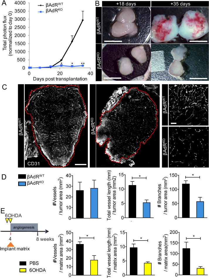
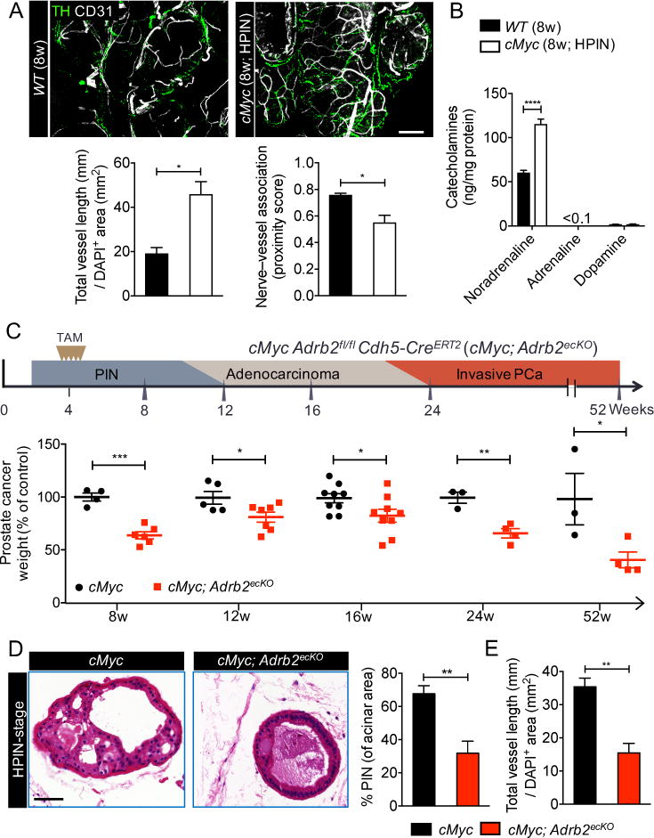
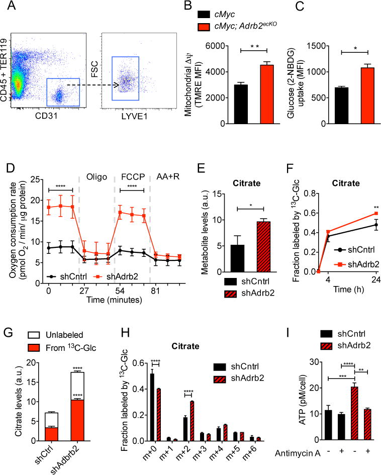
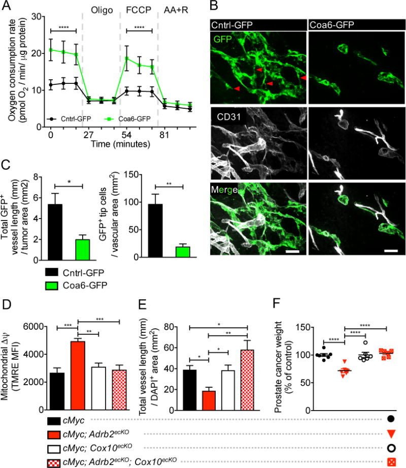
Nerves closely associate with blood vessels and help to pattern the vasculature during development. Recent work suggests that newly formed nerve fibers may regulate the tumor microenvironment, but their exact functions are unclear. Studying mouse models of prostate cancer, we show that endothelial β-adrenergic receptor signaling via adrenergic nerve-derived noradrenaline in the prostate stroma is critical for activation of an angiogenic switch that fuels exponential tumor growth. Mechanistically, this occurs through alteration of endothelial cell metabolism. Endothelial cells typically rely on aerobic glycolysis for angiogenesis. We found that the loss of endothelial Adrb2, the gene encoding the β2-adrenergic receptor, leads to inhibition of angiogenesis through enhancement of endothelial oxidative phosphorylation. Codeletion of Adrb2 and Cox10, a gene encoding a cytochrome IV oxidase assembly factor, prevented the metabolic shift induced by Adrb2 deletion and rescued prostate cancer progression. This cross-talk between nerves and endothelial metabolism could potentially be targeted as an anticancer therapy.


Figures

Fig. 1
Fig. 1. Loss of P-adrenergic signaling in the prostate microenvironment arrests tumor growth and angiogenesis
(A to D) PC-3 cells stably expressing luciferase were orthotopically implanted into 8- week-old Balb/c nu/nu prostates. βAdR = Adrb2 + Adrb3. (A) Tumor growth was monitored in vivo weekly by bioluminescence, n = 6 mice per condition. (B) Comparison of orthotopic tumor size pre (+18 days) and post (+35days) angiogenic switch. Scale bars, 5mm. (C) Immunofluorescence analyses of the vasculature in similar-sized tumors (outlined in red) before the angiogenic switch (+18 days). Cross- sectional montage of prostate xenografts from βAdRWT (left) and βAdRKO (middle) mice, and magnified view of βAdRWT vasculature (right, top) and βAdRKO vasculature (right, bottom). CD31 = vasculature. Montage scale bar, 500μm; magnified-view scale bar, 100μm. (D) Quantification of angiogenesis in orthotopic tumors (vessels traced using Simple Neurite Tracer). n = 4 mice per condition. (E) Experimental design and quantification of vessels recruited into orthotopic type I collagen matrix after sympathectomy with 6-hydroxydopamine (60HDA). PBS = phosphate-buffered saline, n = 4 mice per condition. *P<0.05; **P<0.01. Error bars = indicate SEM.
Fig. 2
Fig. 2. Prostate endothelial cells closely associate with adrenergic nerves and require Adrb2 for cancer progression and angiogenesis
(A) Thick-section images and quantification of the association between adrenergic nerves and the prostate vasculature in the high-grade prostatic intraepithelial neoplasia (HPIN) stage. Representative images of wild-type (WT) prostate (left, top) and HPIN stage prostate (right, top). TH = tyrosine hydroxylase; CD31 = vasculature. Scale bar, 100μm. Quantification of vessel density (left, bottom) and proximity of association between nerves and vessels (right, bottom). n = 4 mice per condition. (B) Catecholamine levels in HPIN-stage prostate quantified by high-performance liquid chromatography. n = 4 mice per condition. (C) Cdh5-CreERT2 deletion of Adrb2 in endothelial cells and its effect at various histopathological stages (schema: top; prostate weight: bottom). TAM = tamoxifen; PCa = prostate cancer. (D and E) Effect of endothelial Adrb2 deletion on HPIN pathology, as shown by (D) representative hematoxylin-and-eosin histology (left). Scale bar, 50μm. Prevalence of PIN (right). n = 4 mice per condition. (E) Quantification of vascular density. n = 4 mice per condition. *P<0.05; **P<0.01; ***P<0.001; ****P<0.0001. Error bars = indicate SEM.
Fig. 3
Fig. 3. Adrb2 depletion increases endothelial oxidative metabolism
(A to C) FACS analysis of HPIN-stage endothelial cells. (A) Representative endothelial isolation plot from prostates. FSC = forward scatter. (B) Quantification of mitochondrial membrane potential (Δψ) by tetramethylrhodamine ethyl ester (TMRE) staining, n = 5 mice per condition. MFI = mean fluorescent intensity. (C) Quantification of endothelial glucose uptake by 2-NBDG. n = 6 or 7 mice per condition. (D to I) Metabolism and ATP production was assessed in shCntrl and shAdrb2 primary prostate endothelial cells after incubation with noradrenaline. (D) Oxygen consumption rates at baseline and in the presence of oligomycin, FCCP [carbonyl cyanide p-(trifl uoromethoxy) phenylhydrazone], and antimycin A + rotenone (AA+R). n = 4 independent experiments. Error bars indicate = SD. (E to H) Different analyses from the same set of experiments, n = 3 replicates per condition per time point. Metabolite levels were normalized to internal standard and to sample protein content. (E) Intracellular levels of the tricarboxylic acid cycle metabolite citrate, a.u., arbitrary units. (F) Total fraction of citrate containing 13C-label from [U-13C]-glucose. (G) Relative intracellular levels of citrate labeling derived from [U-13C]-glucose at 24 hours. (H) Fraction of each isotopologue of citrate (mass isotopologue distribution) after culture in 5mM [U-13C]-glucose for 24 hours. (I) Measurement of intracellular ATP levels in the presence or absence of antimycin A, an inhibitor of the electron transport chain. n = 3 independent experiments. *P<0.05; **P<0.01; ***P<0.001; ****P<0.0001. Error bars = indicate SEM.
Fig. 4
Fig. 4. Increased endothelial oxidative metabolism inhibits angiogenesis, and conditional Cox10 deletion rescues endothelial metabolism, angiogenesis, and cancer progression
(A) Effect of Coa6 overexpression (Coa6-GFP) on oxygen consumption rates at baseline and in the presence of oligomycin, FCCP, and antimycin A + rotenone (AA+R). n = 4 independent experiments. Error bars indicate = SD. (B and C) Immunofluorescent analysis of orthotopically co-transplanted Cntrl-GFP or Coa6-GFP endothelial cells and PC-3 tumor cells to assess in vivo vessel formation. (B) Cntrl-GFP vessels, left; Coa6-GFP vessels, right. GFP = transplanted GFP-tagged endothelial cells; CD31 = vasculature. GFP+ tip cells are indicated by red arrow heads. Scale bars, 50μm. (C) Quantification of vessel density (left) and tip cell formation (right), n = 3 mice per condition. (D to F) HPIN-stage FACS analysis of Δψ (D), vessel density (E), and prostate cancer weight (F) in cMyc, cMyc; Adrb2ecKO, cMyc; Cox10ecKO, and double cMyc; Adrb2ecKO; Cox10ecKO mice, showing restoration of glycolitic metabolism, angiogenesis, and cancer progression after co-deletion of Adrb2 and Cox10 in endothelial cells, n = 6 or 7 mice per condition. Error bars indicate SEM. *P<0.05. **P<0.01. ***P<0.001. ****P<0.0001.

Comment in

References

    1. Folkman J, Watson K, Ingber D, Hanahan D. Induction of angiogenesis during the transition from hyperplasia to neoplasia. Nature. 1989;339:58–61. - PubMed
    1. Bergers G, Benjamin LE. Tumorigenesis and the angiogenic switch. Nature reviews Cancer. 2003;3:401–410. - PubMed
    1. Mukouyama YS, Shin D, Britsch S, Taniguchi M, Anderson DJ. Sensory nerves determine the pattern of arterial differentiation and blood vessel branching in the skin. Cell. 2002;109:693–705. - PubMed
    1. Li W, et al. Peripheral nerve-derived CXCL12 and VEGF-A regulate the patterning of arterial vessel branching in developing limb skin. Developmental cell. 2013;24:359–371. - PMC - PubMed
    1. Ekstrand AJ, et al. Deletion of neuropeptide Y (NPY) 2 receptor in mice results in blockage of NPY-induced angiogenesis and delayed wound healing. Proceedings of the National Academy of Sciences of the United States of America. 2003;100:6033–6038. - PMC - PubMed

Publication types

MeSH terms