. 2017 Oct 20;358(6361):381-386.
doi: 10.1126/science.aan5468.

D4 dopamine receptor high-resolution structures enable the discovery of selective agonists

Affiliations

D4 dopamine receptor high-resolution structures enable the discovery of selective agonists

Sheng Wang et al. Science. .

Abstract

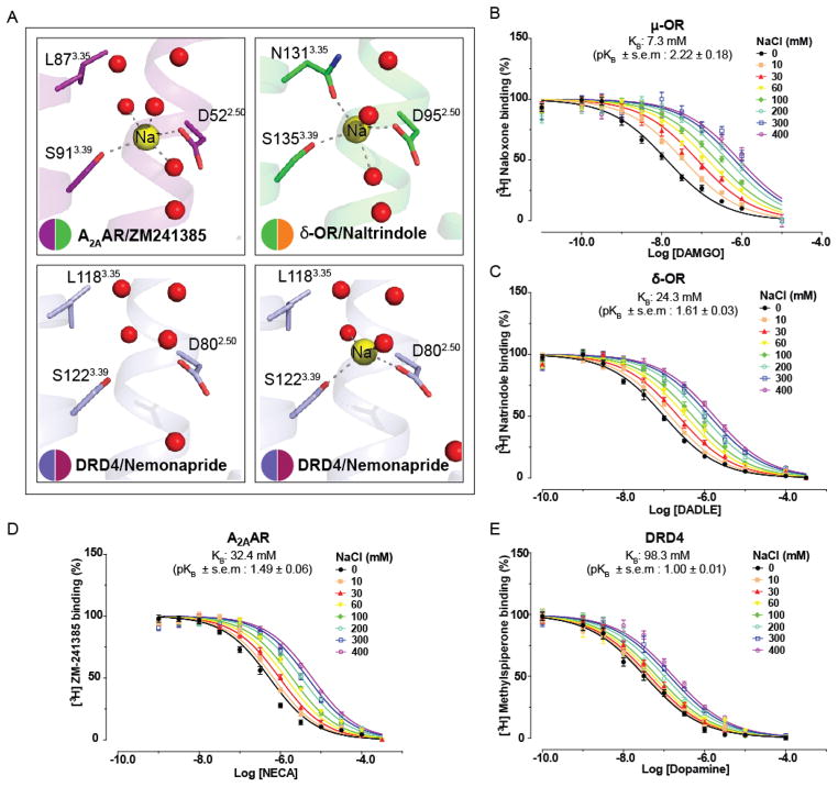
Dopamine receptors are implicated in the pathogenesis and treatment of nearly every neuropsychiatric disorder. Although thousands of drugs interact with these receptors, our molecular understanding of dopaminergic drug selectivity and design remains clouded. To illuminate dopamine receptor structure, function, and ligand recognition, we determined crystal structures of the D4 dopamine receptor in its inactive state bound to the antipsychotic drug nemonapride, with resolutions up to 1.95 angstroms. These structures suggest a mechanism for the control of constitutive signaling, and their unusually high resolution enabled a structure-based campaign for new agonists of the D4 dopamine receptor. The ability to efficiently exploit structure for specific probe discovery-rapidly moving from elucidating receptor structure to discovering previously unrecognized, selective agonists-testifies to the power of structure-based approaches.


Figures

Fig. 1
Fig. 1. Human D4 –nemonapride complex
A, Overview of the DRD4-nemonapride complex structure with water molecules depicted as red spheres. B, Conformation of the binding pocket with nemonapride shown by sticks with magenta carbons. The protein is displayed in cartoon representation, with the 11 contact residues within 4.0 Å from the ligand shown by slate blue sticks. Structured water molecules are shown as red spheres. Ballesteros–Weinstein numbering is shown as superscript. C, Diagram of ligand interactions in the binding pocket side chains at 4.0 Å cut-off. Hydrogen bonds are shown in dashed lines. D, Side views of the sliced binding pocket in DRD4-nemonapride and DRD3-eticlopride complex. The pocket surfaces are colored gray. Ligands are shown as capped sticks with carbons colored magenta (Nemonapride) and orange (eticlopride). E, the different position of phenylalanine in DRD4 (slate blue) and DRD3 (green). Ligands are colored as in panel D.
Fig. 2
Fig. 2. Structural and functional details of the DRD4’s allosteric Na+ site
A, Close ups of A2A adenosine receptor (magenta), δ-OR receptor (green), D4 dopamine receptor without sodium (slate blue) and D4 dopamine receptor with sodium (slate blue) respective Na+ allosteric pockets are shown as inserts. The receptors are shown as a ribbon, and residues lining the Na+ cavity are shown as sticks and labeled according to Ballesteros-Weinstein scheme. Water molecules in the cluster are shown as red spheres. Na+ is displayed as yellow spheres. The salt bridge between Na+ and D2.50 and hydrogen bonds are shown as grey dotted lines. B, C, D, E, The allosteric effect of graded dose of sodium on DAMGO, DADLE, NECA or dopamine affinity were respectively measured at μ-OR (B), δ-OR (C), A2AAR (D) and DRD4 (E).
Fig. 3
Fig. 3. Discovery of a new DRD4 selective agonist
A, Structure-based strategy for discovery of novel DRD4 chemotypes from docking screens against large virtual libraries. B, Single-point competition binding assay of 10 candidate molecules against the DRD4 antagonist 3H-N-Methylspiperone. Each ligand was tested at 5 μM and for those with > 50% inhibition affinity was calculated in full displacement curves; data represent mean ± s.e.m. (n = 3 measurements). C, D Docking poses of compounds 3 (C) and 9 (D), new DRD4 agonists. The D4 dopamine receptor and residues are shown in tan. Compounds are shown as capped sticks with carbons colored cyan (compound 3) and green (compound 9). Ballesteros–Weinstein numbering is shown as superscript.
Fig. 4
Fig. 4. Structure-guided optimization of potent DRD4 agonists
A, Overview of structure-guided analog-by-catalog optimization towards compound 9-6-24 (see main text for details). B, C, Docking poses of compounds 9-6 (B) and 9-6-24 (C). Both optimized analogs have a simplified right hand linker while maintaining key interactions and occupying the EBP. The D4 dopamine receptor and residues are shown in tan. Compounds are shown as capped sticks with carbons colored pink (compound 9-6) and green (compound 9-6-24). Ballesteros–Weinstein numbering is shown as superscript. D, E, Normalized concentration-response studies for compound 9-6 (D) and 9-6-24 (E) at human cloned DRD4–mediated activation of Gαi/o and arrestin translocation. Error bars in (D) and (E) denote SEM from a minimum of three assays. See METHODS for details on bias factor calculation. F, G, Compound 9-6 (F) and Compound 9-6-24 (G) were screened against 320 non-olfactory GPCRs for agonism in the arrestin recruitment TANGO assay. Each point shows luminescence normalized to basal level at a given GPCR, with vertical lines indicating the standard error of the mean.

References

    1. Missale C, Nash SR, Robinson SW, Jaber M, Caron MG. Physiol Rev. 1998;78:189–225. - PubMed
    1. Beaulieu JM, Espinoza S, Gainetdinov RR. Br J Pharmacol. 2015;172:1–23. - PMC - PubMed
    1. Chien EY, et al. Science. 2010;330:1091–1095. - PMC - PubMed
    1. Chu R, et al. J Mol Biol. 2002;323:253–262. - PubMed
    1. Rasmussen SG, et al. Nature. 2011;477:549–555. - PMC - PubMed

Publication types

MeSH terms