. 2017 Oct 5:11:311.
doi: 10.3389/fncel.2017.00311. eCollection 2017.

Exacerbation of Brain Injury by Post-Stroke Exercise Is Contingent Upon Exercise Initiation Timing

Affiliations

Exacerbation of Brain Injury by Post-Stroke Exercise Is Contingent Upon Exercise Initiation Timing

Fengwu Li et al. Front Cell Neurosci. .

Abstract

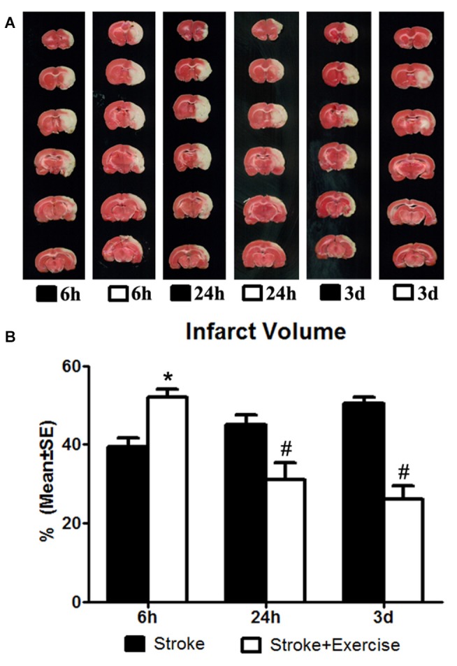
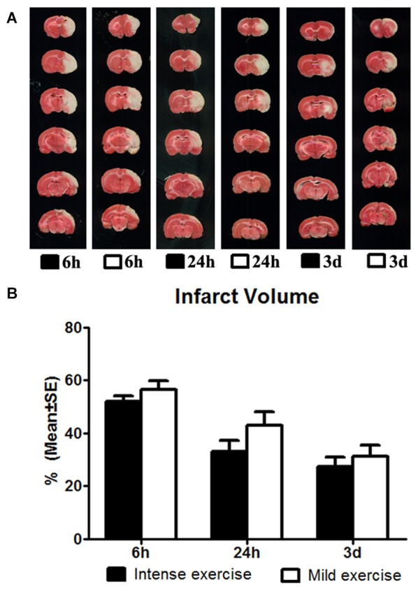
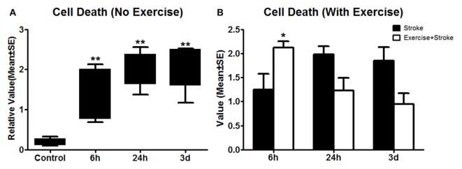
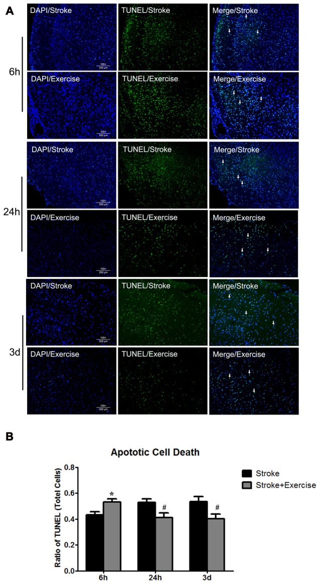
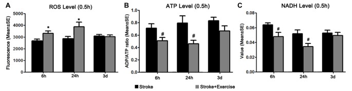
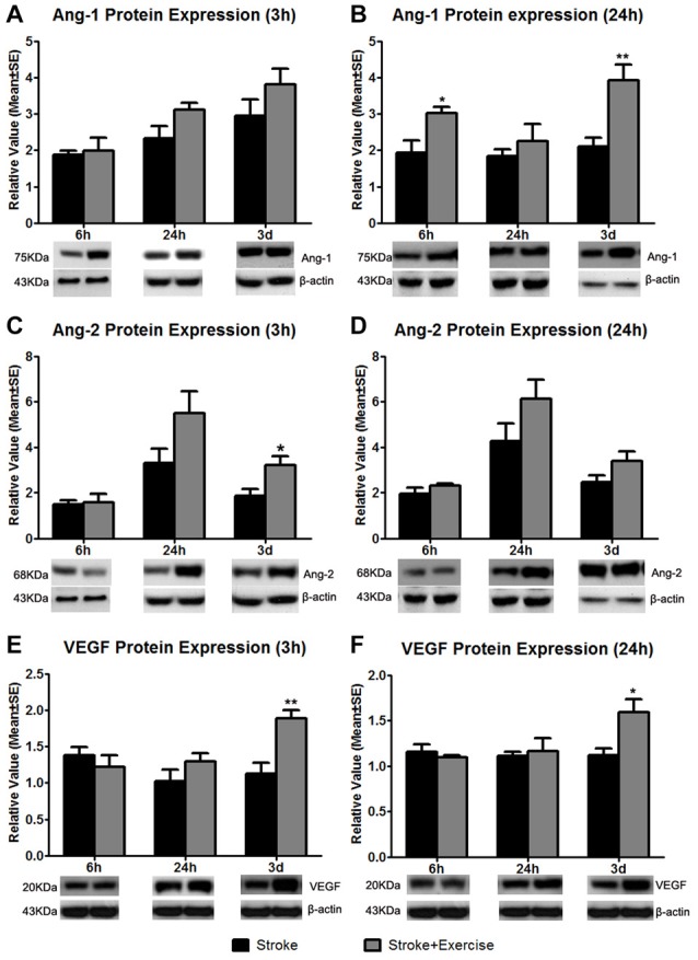
Accumulating evidence has demonstrated that post-stroke physical rehabilitation may reduce morbidity. The effectiveness of post-stroke exercise, however, appears to be contingent upon exercise initiation. This study assessed the hypothesis that very early exercise exacerbates brain injury, induces reactive oxygen species (ROS) generation, and promotes energy failure. A total of 230 adult male Sprague-Dawley rats were subjected to middle cerebral artery (MCA) occlusion for 2 h, and randomized into eight groups, including two sham injury control groups, three non-exercise and three exercise groups. Exercise was initiated after 6 h, 24 h and 3 days of reperfusion. Twenty-four hours after completion of exercise (and at corresponding time points in non-exercise controls), infarct volumes and apoptotic cell death were examined. Early brain oxidative metabolism was quantified by examining ROS, ATP and NADH levels 0.5 h after completion of exercise. Furthermore, protein expressions of angiogenic growth factors were measured in order to determine whether post-stroke angiogenesis played a role in rehabilitation. As expected, ischemic stroke resulted in brain infarction, apoptotic cell death and ROS generation, and diminished NADH and ATP production. Infarct volumes and apoptotic cell death were enhanced (p < 0.05) by exercise that was initiated after 6 h of reperfusion, but decreased by late exercise (24 h, 3 days). This exacerbated brain injury at 6 h was associated with increased ROS levels (p < 0.05), and decreased (p < 0.05) NADH and ATP levels. In conclusion, very early exercise aggravated brain damage, and early exercise-induced energy failure with ROS generation may underlie the exacerbation of brain injury. These results shed light on the manner in which exercise initiation timing may affect post-stroke rehabilitation.

Keywords: ATP; NAD; apoptosis; brain metabolism; infarction; reactive oxygen species.


Figures

Figure 1
Figure 1
(A) 2,3,5-Triphenyltetrazolium chloride (TTC) staining illustrating infarct volumes in ischemic rats (subjected to 2-h middle cerebral artery (MCA) occlusion) with or without exercise initiated at 6 h, 24 h and 3 days after reperfusion. (B) In contrast to the non-exercise groups, very early exercise at 6 h largely (*p < 0.05) increased neural damage, while the later exercise groups conducted at 24 h and 3 days after reperfusion demonstrated a significant (#p < 0.05) reduction in apoptosis.
Figure 2
Figure 2
(A) Comparison of mild and intense exercise (initiated at 6 h, 24 h and 3 days of reperfusion) on infarct volume determined by TTC. (B) Compared to the intense exercise groups, infarct volumes of the mild exercise groups were slightly but not significantly increased after all exercise time points.
Figure 3
Figure 3
(A) Apoptotic cell death detected using ELISA was measured 24 h after exercise termination or the equivalent time point in non-exercise animals. One-way ANOVA of non-exercise groups indicated a significant (**p < 0.01) increase in all non-exercise ischemic rat groups (6 h, 24 h and 3 day) compared to sham-operation controls. (B) In contrast to non-exercise groups, The student t-test indicates that very early exercise initiated at 6 h of reperfusion significantly (*p < 0.05) increased cell death, while groups in which exercise was initiated after 24 h and 3 days of reperfusion exhibited non-significant reductions in apoptosis.
Figure 4
Figure 4
(A) TUNEL of non-exercise and exercise groups (initiated at 6 h, 24 h and 3 days of reperfusion). (B) Compared to stroke groups without exercise, apoptotic cell death was significantly (*p < 0.05) increased after very early exercise (initiated at 6 h of reperfusion), while later exercise (24 h and 3 days) significantly (#p < 0.05) reduced cell death as compared to non-exercise stroke group.
Figure 5
Figure 5
(A) Reactive oxygen species (ROS) levels were examined 0.5 h after exercise termination in ischemic rats with exercise and at equivalent time points in ischemic rats without exercise. Compared to sham controls (reference as 1, not shown), ischemic animals exhibited significantly increased ROS levels. Further increases in ROS production were observed in the early (6 h and 24 h, *p < 0.05) exercise groups as compared to non-exercise controls. Late exercise (initiated after 3 days of reperfusion) did not have a significant impact on ROS levels, as compared to non-exercise ischemic rats. (B) ATP levels were examined 0.5 h after exercise termination in exercise groups and at equivalent time points in non-exercise controls. MCA occlusion significantly decreased ATP production as compared to sham controls (reference as 1, not shown). Early exercise groups (6 h and 24 h, #p < 0.05), though not the late exercise group (3 days), exhibited further reductions in ATP levels as compared to non-exercise controls. (C) NADH levels, measured 0.5 h after exercise termination in exercise groups and at equivalent time points in ischemic rats, were significantly decreased in all non-exercise ischemic groups as compared to sham control (reference as 1, not shown). Exercise initiated after 6 h and 24 h of reperfusion further reduced (#p < 0.05) NADH levels as compared to corresponding non-exercise groups.
Figure 6
Figure 6
Based on sham control (reference as 1, not shown), angiopoietin-1 (Ang-1) expression at 3 h after exercise termination was slightly increased in exercise groups compared to non-exercise ischemic rat groups at 6 h, 24 h and 3 days after reperfusion (A). A significantly enhanced increase in Ang-1 protein expression at 24 h after exercise termination was observed in the very early (6 h; *p < 0.05) and late (3 days; **p < 0.01) exercise groups while a slight increase was seen in the 24 h exercise group (B). Angiopoietin-2 (Ang-2) protein expression was measured 3 and 24 h after exercise termination or the equivalent period (C,D). At 3 h, late exercise (3 days) resulted in a significant (*p < 0.05) increase in Ang-2 protein expression compared to non-exercise ischemic rat groups, while no changes were seen with early exercise (6 and 24 h). For 24 h after exercise termination, exercise at three time points resulted in an increase in Ang-2 protein expression, although they did not reach a significant level. (E) Vascular endothelial growth factor (VEGF) protein expression was measured 3 h after exercise termination or the equivalent period. Only late exercise (3 days) significantly (**p < 0.01) increased VEGF protein expression compared to non-exercise ischemic rat groups. (F) Similar results were seen in which late (3 days) resulted in a significant (*p < 0.05) increase in VEGF protein expression. Representative immunoblots are presented.

References

    1. Almeida A., Moncada S., Bolaños J. P. (2004). Nitric oxide switches on glycolysis through the AMP protein kinase and 6-phosphofructo-2-kinase pathway. Nat. Cell Biol. 6, 45–51. 10.1038/ncb1080 - DOI - PubMed
    1. Arya K. N., Pandian S., Verma R., Garg R. K. (2011). Movement therapy induced neural reorganization and motor recovery in stroke: a review. J. Bodyw. Mov. Ther. 15, 528–537. 10.1016/j.jbmt.2011.01.023 - DOI - PubMed
    1. Austin M. W., Ploughman M., Glynn L., Corbett D. (2014). Aerobic exercise effects on neuroprotection and brain repair following stroke: a systematic review and perspective. Neurosci. Res. 87, 8–15. 10.1016/j.neures.2014.06.007 - DOI - PubMed
    1. Bernhardt J., Dewey H., Collier J., Thrift A., Lindley R., Moodie M., et al. . (2006). A very early rehabilitation trial (AVERT). Int. J. Stroke 1, 169–171. 10.1111/j.1747-4949.2006.00044.x - DOI - PubMed
    1. Bernhardt J., Langhorne P., Lindley R. I., Thrift A. G., Ellery F., Collier J., et al. . (2015). Efficacy and safety of very early mobilisation within 24 h of stroke onset (AVERT): a randomised controlled trial. Lancet 386, 46–55. 10.1016/s0140-6736(15)60690-0 - DOI - PubMed