. 2017 Oct;10(10):761-771.
doi: 10.1177/1756283X17725998. Epub 2017 Aug 25.

Increased expression of hepatocyte nuclear factor 4 alpha transcribed by promoter 2 indicates a poor prognosis in hepatocellular carcinoma

Affiliations

Increased expression of hepatocyte nuclear factor 4 alpha transcribed by promoter 2 indicates a poor prognosis in hepatocellular carcinoma

Shao-Hang Cai et al. Therap Adv Gastroenterol. 2017 Oct.

Abstract

Background: Hepatocyte nuclear factor 4 alpha (HNF4α) plays an important role in tumourigenesis. There is growing evidence indicating that HNF4α transcribed by promoter 1 (P1-HNF4α) is expressed at relatively low levels in HCC and its presence predicts a favourable outcome for hepatocellular carcinoma (HCC) patients. However, the role of HNF4α transcribed by promoter 2 (P2-HNF4α) in HCC remains unclear.

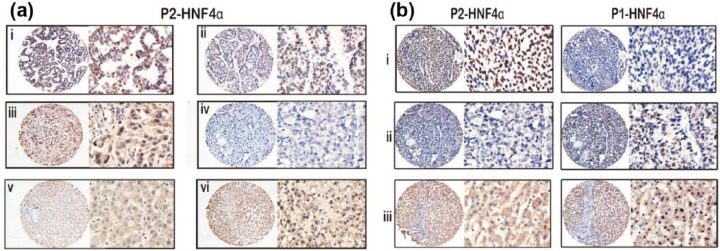
Methods: A total of 615 HCC specimens were obtained to construct tissue microarrays and perform immunohistochemistry. The relationship between P2-HNF4α and clinical features of HCC patients were analysed. Kaplan-Meier analysis was conducted to assess the prognostic value of P2-HNF4α.

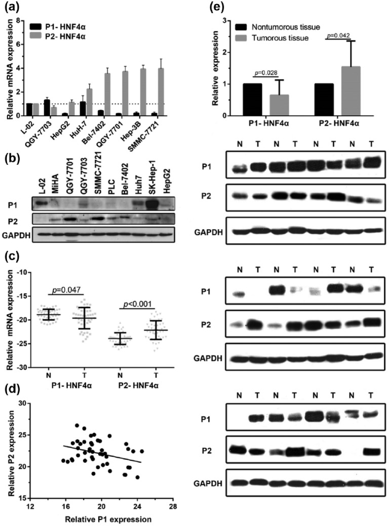
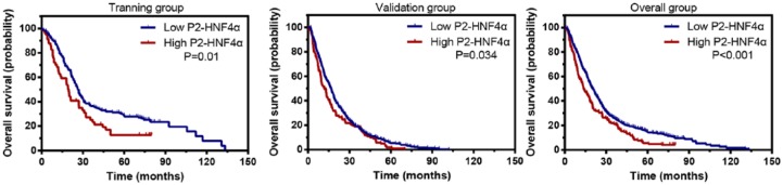
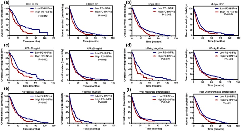
Results: The results showed that the expression of P2-HNF4α in HCC was noticeably increased in HCC tissues compared with the nontumourous tissues. In addition, P1-HNF4α expression was negatively correlated with P2-HNF4α expression (p = 0.023). High P2-HNF4α expression was significantly associated with poor differentiation of HCC (p = 0.002) and vascular invasion (p = 0.017). Kaplan-Meier analysis showed that P2-HNF4α expression was closely correlated with overall survival in the training group (p = 0.01), validation group (p = 0.034), and overall group of patients with HCC (p < 0.001).

Conclusions: Our data show that the role of HNF4α in cancer development needs to be further refined. P2-HNF4α, different from P1-HNF4α, is markedly upregulated and serves as an oncogene-associated protein in HCC. Our study therefore provides a promising biomarker for prognostic prediction and a potential therapeutic target for HCC.

Keywords: hepatocellular carcinoma; hepatocyte nuclear factor 4α; prognostic biomarker; promoter 1; promoter 2.


Conflict of interest statement

Conflict of interest statement: The authors declare that there is no conflict of interest.

Figures

Figure 1.
Figure 1.
P1-HNFα expression is decreased and P2-HNF4α expression is increased in HCC cell lines by polymerase chain reaction and western blotting (a–b). mRNA and protein levels of P1- and P2-HNF4α were measured in fresh HCC tissues (T) and corresponding adjacent nontumourous tissues (N) (c and e). Correlation of P1- and P2-HNF4α mRNA expression (r = −0.339, p = 0.023; d). Immortalized hepatocytes: L-02, MiHA; HCC cell lines: QGY-7703, HepG2, Huh-7, Bel-7402, QGY-7701, Hep3B, SMMC-7721, PLC, and SK-Hep-1. HCC, hepatocellular carcinoma; HNF4α, hepatocyte nuclear factor 4 alpha; P1, promoter 1; P2, promoter 2.
Figure 2.
Figure 2.
P2-HNF4α expression is increased in HCC samples as shown by immunohistochemistry. Representative images of strong (ai), moderate (aii), weak (aiii), and negative (aiv) immunoreactivities of P2-HNF4α in HCC samples, as well as negative (av) and positive (avi) staining of P2-HNF4α in normal liver tissues are shown (left panel: magnification ×100; right panel: magnification ×400). (b) P2-HNF4α positive expressing HCC tissue is always accompanied by negative P1-HNF4α expression (bi). Negative expression of P2-HNF4α in HCC tissue is accompanied by positive P1-HNF4α expression (bii). Negative expression of P2-HNF4α in nontumourous tissue is accompanied by positive P1-HNF4α expression (biii). HCC, hepatocellular carcinoma; HNF4α, hepatocyte nuclear factor 4 alpha; P1, promoter 1; P2, promoter 2.
Figure 3.
Figure 3.
Representative images of well-differentiated (a), moderately-differentiated (b) and poorly-differentiated HCC (c) in liver biopsy tissue. In the HCC samples with better differentiation, the proportion of cells with high expression of P1-HNF4α is significantly higher than P2-HNF4α, whereas in HCC samples with poor differentiation, P2-HNF4α expression was more common. HCC, hepatocellular carcinoma; HNF4α, hepatocyte nuclear factor 4 alpha; P1, promoter 1; P2, promoter 2.
Figure 4.
Figure 4.
High P2-HNF4α expression is correlated with an unfavourable prognosis in both the training and validation cohorts. Kaplan–Meier analysis shows the significant differences in overall survival between postoperative HCC patients with high and low P2-HNF4α expression in training (n = 241), validation (n = 374), and overall cohorts. HCC, hepatocellular carcinoma; HNF4α, hepatocyte nuclear factor 4 alpha; P2, promoter 2.
Figure 5.
Figure 5.
High P2-HNF4α expression is associated with unfavourable outcomes in subgroups of HCC patients. Stratified survival analyses show that P2- HNF4α expression is correlated with overall survival in small and large HCCs (a) in single and multinodular HCC (b), in HCCs with normal and abnormal AFP levels (c), in HBsAg positive and negative HCCs (d), in HCCs with or without microvascular invasion (e), and in HCCs with well-moderate differentiation and poor-undifferentiated differentiation (f). HBsAg, hepatitis B surface antigen; HCC, hepatocellular carcinoma; HNF4α, hepatocyte nuclear factor 4 alpha; P1, promoter 1; P2, promoter 2.

References

    1. Babeu JP, Boudreau F. Hepatocyte nuclear factor 4-alpha involvement in liver and intestinal inflammatory networks. World J Gastroenterol 2014; 20: 22–30. - PMC - PubMed
    1. Hanawa M, Takayama K, Sakurai F, et al. Hepatocyte nuclear factor 4 alpha promotes definitive endoderm differentiation from human induced pluripotent stem cells. Stem Cell Rev 2016; 13: 542–551. - PubMed
    1. Chen WS, Manova K, Weinstein DC, et al. Disruption of the HNF-4 gene, expressed in visceral endoderm, leads to cell death in embryonic ectoderm and impaired gastrulation of mouse embryos. Genes Dev 1994; 8: 2466–2477. - PubMed
    1. Drewes T, Senkel S, Holewa B, et al. Human hepatocyte nuclear factor 4 isoforms are encoded by distinct and differentially expressed genes. Mol Cell Biol 1996; 16: 925–931. - PMC - PubMed
    1. Torres-Padilla ME, Fougere-Deschatrette C, Weiss MC. Expression of HNF4alpha isoforms in mouse liver development is regulated by sequential promoter usage and constitutive 3’ end splicing. Mech Dev 2001; 109: 183–193. - PubMed

LinkOut - more resources