. 2017 Jul 8;4(10):1700034.
doi: 10.1002/advs.201700034. eCollection 2017 Oct.

Functional Recovery of Contused Spinal Cord in Rat with the Injection of Optimal-Dosed Cerium Oxide Nanoparticles

Affiliations

Functional Recovery of Contused Spinal Cord in Rat with the Injection of Optimal-Dosed Cerium Oxide Nanoparticles

Jong-Wan Kim et al. Adv Sci (Weinh). .

Abstract

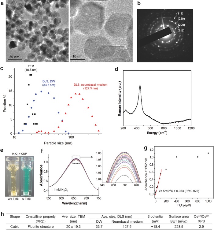
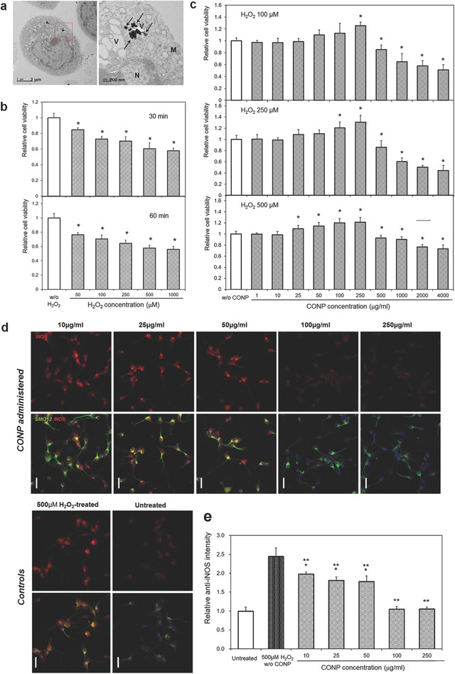
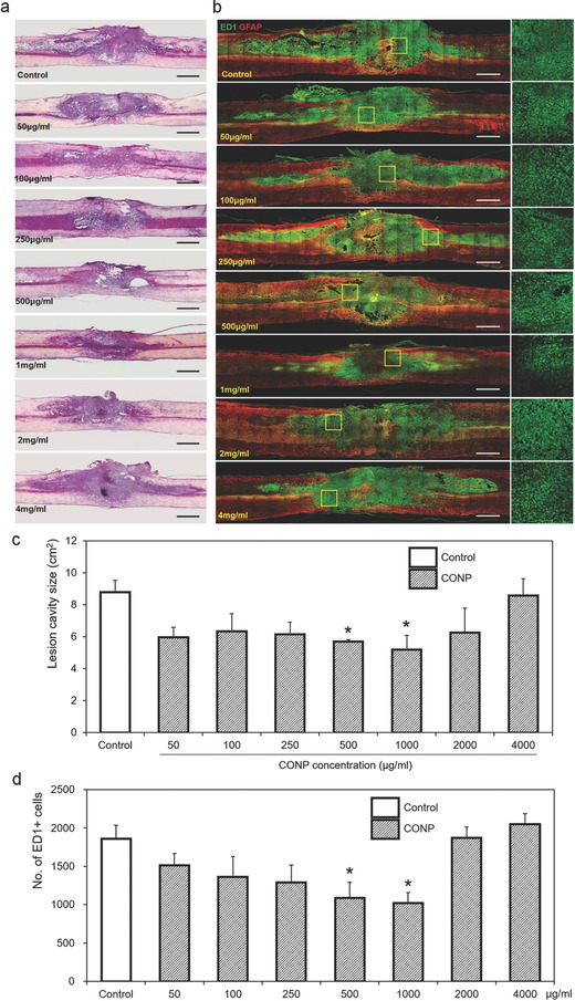
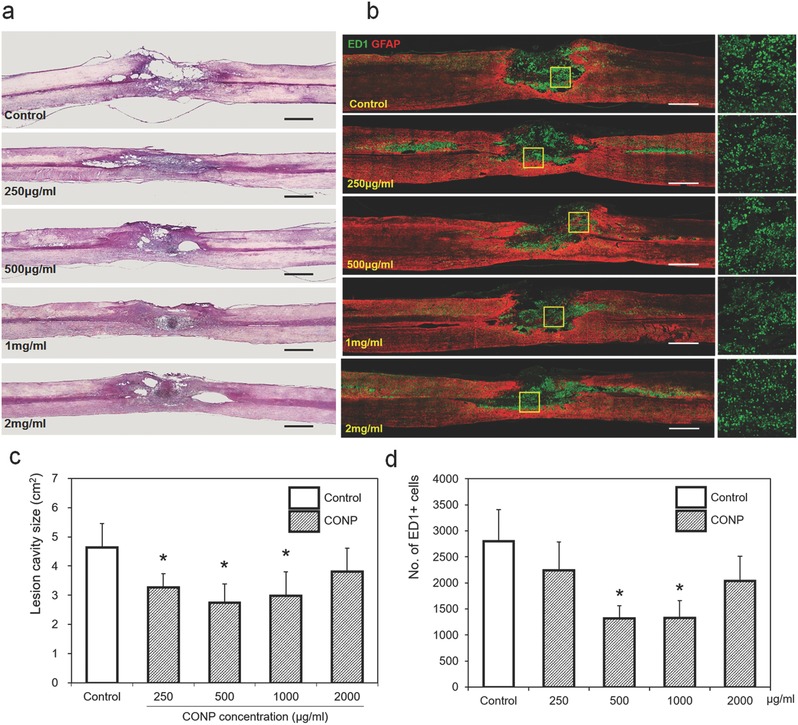
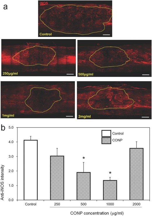
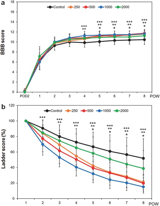
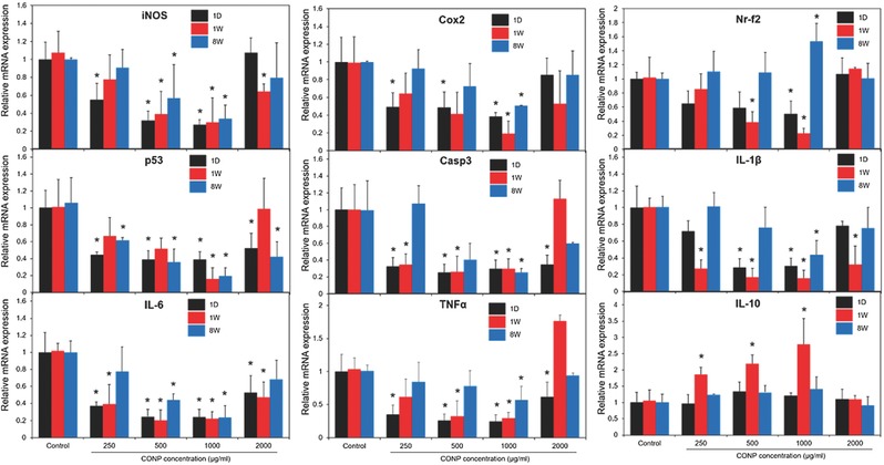
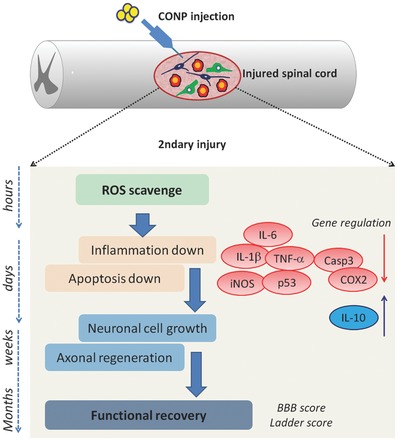
Spinal cord injury (SCI) produces excess reactive oxygen species (ROS) that can exacerbate secondary injury and lead to permanent functional impairment. Hypothesizing that cerium oxide nanoparticles (CONPs) as an effective ROS scavenger may offset this damaging effect, it is first demonstrated in vitro that CONPs suppressed inducible nitric oxide synthase (iNOS) generation and enhanced cell viability of hydrogen peroxide (H2O2)-insulted cortical neurons. Next, CONPs are administered at various does (50-4000 µg mL-1) to a contused spinal cord rat model and monitored the disease progression for up to eight weeks. At one day postinjury, the number of iNOS+ cells decreases in the treated groups compared with the control. At one week, the cavity size and inflammatory cells are substantially reduced, and the expression of proinflammatory and apoptotic molecules is downregulated with a concurrent upregulation of anti-inflammatory cytokine. By eight weeks, the treated groups show significantly improved locomotor functions compared with the control. This study shows for the first time that injection of optimal-dosed CONPs alone into contusion-injured spinal cord of rats can reduce ROS level, attenuate inflammation and apoptosis, and consequently help locomotor functional recovery, adding a promising and complementary strategy to the other treatments of acute SCI.

Keywords: anti‐inflammation; cerium oxide nanoparticles; functional recovery; reactive oxygen species; spinal cord injury.


Figures

Figure 1
Figure 1
Characteristics of cerium oxide nanoparticles (CONPs). a) TEM images of CONPs at low and high magnification, and b) selected area diffraction pattern of the crystal. c) Size distribution of CONPs, calculated from TEM images and also measured by DLS (in DW or neurobasal medium). d) Raman spectrum. e–g) Oxidase‐like activity of CONPs, evaluated by monitoring the redox reaction between TMB and H2O2 in the presence of the CONPs; optical view showing a color change by the reaction (e), UV–vis spectroscopic intensity measured time‐dependently (20 cycles per 5 min) at a broad wavelength scan using 1 × 10⁻³ m H2O2 (f) and then recorded at a specific peak 652 nm with varying H2O2 dose up to 1000 µm (showing a linear relationship up to ≈100 µm) (g). h) Summary of properties including shape, size (by TEM and DLS method), ζ‐potential, surface area (by BET), and Ce atomic oxidation status (Ce4+/Ce3+, by XPS).13
Figure 2
Figure 2
a) CONP (100 µg mL−1) internalization to H2O2 (500 × 10−6 m)‐insulted cortical neurons, visualized by TEM; right is a magnified image of a red box in left. Black arrows indicate CONPs (N: nucleus, M: mitochondria, V: vesicle). b–e) Effects of CONP administration to H2O2‐modeled cortical neurons in vitro: Neuronal viability, after H2O2 treatment at varying doses of 50 × 10−6 m to 1000 × 10−6 m for 30 or 60 min, is reduced dose‐dependently (b); *p < 0.05 compared with control by Mann–Whitney U test. Administration of CONP at varying concentrations of 1–4000 µg mL−1 to the H2O2‐insulted neurons recovers the cell viability (c); H2O2 varied at 100 × 10−6 m, 250 × 10−6 m, or 500 × 10−6 m; *p < 0.05 compared with control by Mann–Whitney U test. In vitro iNOS generation assay (d, e); representative images of anti‐iNOS (red) and anti‐SMI312 (green) positive cortical neurons (scale bar = 20 µm), and the relative intensity of anti‐iNOS levels following 500 × 10−6 m H2O2‐treatment and the concomitant application of various concentrations of CONPs; *p < 0.05 compared with untreated control group, and **p < 0.05 compared with H2O2‐treated group, by Kruskal–Wallis test with Bonferroni correction.
Figure 3
Figure 3
Representative a) H&E and b) immunohistochemical images of the injured spinal cord one week after the administration of CONPs with different concentrations from 50 to 4000 µg mL−1. The yellow boxes magnified on right side. c) Size of lesion cavity measured from the sagittal images of H&E staining, and d) number of ED1‐positive inflammatory cells calculated from the sagittal images of immunohistochemistry. Scale bar = 1 mm. *p < 0.05 compared with control by Mann–Whitney U test.
Figure 4
Figure 4
Representative a) H&E and b) immunohistochemical images of the injured spinal cord eight weeks after the application of CONPs with different concentrations from 250 to 2000 µg mL−1. The yellow boxes magnified on right side. c) Size of lesion cavity measured from the sagittal images of H&E staining, and d) number of ED1‐positive inflammatory cells calculated from the sagittal images of immunohistochemistry. Scale bar = 1 mm. *p < 0.05 compared with control by Mann–Whitney U test.
Figure 5
Figure 5
In vivo iNOS generation assay. a) Represent images of anti‐iNOS positive cells within injured spinal cord 24 h after the SCI. The lesion cavity outlined by yellow dots. b) Relative intensity of anti‐iNOS positive cells quantified. Scale bar = 500 µm. *p < 0.05 compared with control by Mann–Whitney U test.
Figure 6
Figure 6
Locomotor functions of spinal cord injured rats after the application of CONPs until eight weeks. a) BBB score, and b) ladder score. *p < 0.05 between 250 µg mL−1 and control, **p < 0.05 between 500 µg mL−1 and control, and ***p < 0.05 between 1000 µg mL−1 and control, by Kruskal–Wallis test with Bonferroni correction.
Figure 7
Figure 7
In vivo mRNA expression levels of genes associated with anti‐inflammatory and apoptosis, including ROS, Cox2, Nr‐f2, p53, Casp3, IL‐1β, IL‐6, TNFα, and IL‐10 within the injured spinal cord treated with CONPs at different concentrations (250–2000 µg mL−1), measured at one day, one week, and eight weeks postinjury. *p < 0.05 compared with control at the same period by Mann–Whitney U test.
Figure 8
Figure 8
Schematic illustration showing the series of events involved in secondary injury after spinal cord contusion and the therapeutic regulation of directly injected CONPs through ROS scavenging, suppressing inflammation and apoptosis, enhancing neuronal cell growth and axonal regeneration, and consequently functional recovery.

References

    1. Ho C. H., Wuermser L. A., Priebe M. M., Chiodo A. E., Scelza W. M., Kirshblum S. C., Arch. Phys. Med. Rehabil. 2007, 88, S49. - PubMed
    1. Silva N. A., Sousa N., Reis R. L., Salgado A. J., Prog. Neurobiol. 2014, 114, 25. - PubMed
    1. Schwab J. M., Zhang Y., Kopp M. A., Brommer B., Popovich P. G., Exp. Neurol. 2014, 258, 121. - PMC - PubMed
    1. a) Bao F., Liu D., Neuroscience 2004, 126, 285; - PubMed
    2. b) Gwak Y. S., Hassler S. E., Hulsebosch C. E., Pain 2013, 154, 1699; - PubMed
    3. c) Plemel J. R., Wee Yong V., Stirling D. P., Exp. Neurol. 2014, 258, 91. - PubMed
    1. a) Liu D., Bao F., Neuroscience 2015, 285, 81; - PMC - PubMed
    2. b) Lee J. Y., Maeng S., Kang S. R., Choi H. Y., Oh T. H., Ju B. G., Yune T. Y., J. Neurotrauma 2014, 31, 582. - PMC - PubMed