. 2018:1675:183-201.
doi: 10.1007/978-1-4939-7318-7_12.

Identification of Open Chromatin Regions in Plant Genomes Using ATAC-Seq

Affiliations

Identification of Open Chromatin Regions in Plant Genomes Using ATAC-Seq

Marko Bajic et al. Methods Mol Biol. 2018.

Abstract

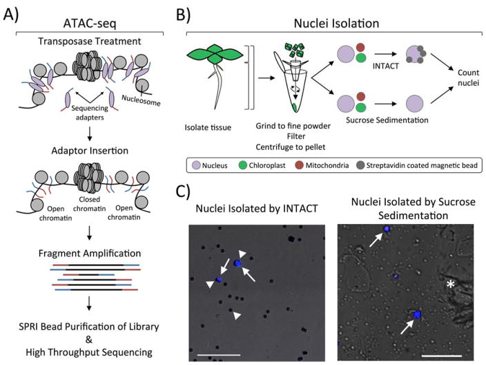
Identifying and characterizing highly accessible chromatin regions assists in determining the location of genomic regulatory elements and understanding transcriptional regulation. In this chapter, we describe an approach to map accessible chromatin features in plants using the Assay for Transposase-Accessible Chromatin, combined with high-throughput sequencing (ATAC-seq), which was originally developed for cultured animal cells. This technique utilizes a hyperactive Tn5 transposase to cause DNA cleavage and simultaneous insertion of sequencing adapters into open chromatin regions of the input nuclei. The application of ATAC-seq to plant tissue has been challenging due to the difficulty of isolating nuclei sufficiently free of interfering organellar DNA. Here we present two different approaches to purify plant nuclei for ATAC-seq: the INTACT method (Isolation of Nuclei TAgged in specific Cell Types) to isolate nuclei from individual cell types of the plant, and tissue lysis followed by sucrose sedimentation to isolate sufficiently pure total nuclei. We provide detailed instructions for transposase treatment of nuclei isolated using either approach, as well as subsequent preparation of ATAC-seq libraries. Sequencing-ready ATAC-seq libraries can be prepared from plant tissue in as little as one day. The procedures described here are optimized for Arabidopsis thaliana but can also be applied to other plant species.

Keywords: ATAC-seq; Chromatin; Enhancer; INTACT system; Nucleosome; Nucleus; Transcription factor; Transposition.


Figures

Figure 1
Figure 1. ATAC-seq profiling using nuclei isolated by INTACT or sucrose sedimentation
A) Overview of the ATC-seq procedure. Nuclei are incubated with sequencing adapter-loaded Tn5 transposase, which diffuses into the nucleus to interact with chromatin. Sequencing adapters are inserted into open chromatin regions, and the fragmented DNA is amplified wherever the sequencing adapters were inserted. This generates a library of DNA fragments in which each end represents an insertion site. The amplified libraries are purified and sequenced with next generation sequencing. B) Two different methods for purifying nuclei from Arabidopsis thaliana can be used: 1) INTACT for isolating nuclei from specific cell types, and 2) sucrose sedimentation to isolate total nuclei from input tissue. The two methods have the same initial steps: tissue is collected from a specific part of the plant (root, leaf, or the entire plant), ground to a fine powder, resuspended, filtered, and centrifuged to pellet nuclei and cellular debris. Nuclei isolation using tissue that expresses INTACT transgenes uses streptavidin coated magnetic beads to affinity purify biotinylated nuclei out of the resuspended pellet. This allows for the isolation of nuclei from specific cell-types that express the nuclear tagging fusion (NTF) and the biotin ligase BirA, resulting in very low contamination by organellar genomes. Alternatively, total nuclei can be isolated from tissue by resuspending the nuclei/debris pellet in a buffer with Triton X-100 to lyse organelles and centrifuging through a dense sucrose layer. Nuclei isolated from both procedures are stained with DAPI and quantified using a hemocytometer. C) Fluorescent microscope images of nuclei (white arrows) stained with the DNA-binding dye DAPI (blue) isolated either through INTACT or sucrose sedimentation. INTACT isolated nuclei are identified by their DAPI-fluorescence and binding to multiple beads (white arrowhead). Beads are easily visualized by increasing transmission of white light while viewing the nuclei in the DAPI channel. Sucrose sedimentation isolated nuclei (white arrows) are DAPI-stained objects around 4–6 μm in diameter, although they can vary in size and shape depending on starting tissue. Much more cellular debris (white asterisk) is observed in sucrose sedimentation-isolated nuclei as compared to INTACT-purified nuclei, but this should not impact the procedure described here. Each picture contains a 50 μm scale bar shown at the bottom left.
Figure 2
Figure 2. ATAC-seq library preparation and high-throughput sequencing
A) An amplified ATAC-seq library purified with Ampure XP beads (lane “1”) was resolved in a 2% agarose gel stained with ethidium bromide. Lane “M” is the molecular weight marker lane. Amplified library fragments generally range in size from 180 bp to several kb in size. The size distribution of the resolved gel may vary somewhat, but the final product should be free of adapter dimers (distinct band around 125 bp) and primer dimers (distinct band around 80 bp). See Note 11. B) Insert sizes of ATAC-seq paired-end reads from 50,000 nuclei isolated by INTACT from non-hair cells calculated using the InsertSizeMetrics option from Picard Tools (Note 13). The distribution shows periodicity of helical pitch of DNA for fragments smaller than 200 bp. Fragments containing one or more nucleosomes, related to insert periodicity increasing in 150 bp, were not observed using the transposase:nuclei and bead:DNA ratios described in this protocol. C) Integrated Genome Viewer snapshot of four different libraries sequenced on the Illumina platform. The tracks shown are of ATAC sequencing reads from INTACT isolated nuclei from root hair cells (orange), root non-hair cells (purple), root tip (cyan), and sucrose sedimentation isolated nuclei from 1 cm root tip (navy). Gene tracks are shown below the ATAC-seq tracks and a 25 kb scale bar is show

References

    1. Li B, Carey M, Workman JL. The role of chromatin during transcription. Cell. 2007;128:707–719. - PubMed
    1. Song L, Crawford GE. DNase-seq: a high-resolution technique for mapping active gene regulatory elements across the genome from mammalian cells. Cold Spring Harb Protoc. 2010;2010 doi: 10.1101/pdb.prot5384. pdb prot5384. - DOI - PMC - PubMed
    1. Ken Z. Micrococcal Nuclease Analysis of Chromatin Structure. Current Protocols in Molecular Biology. 2005;21(1):1–17. - PubMed
    1. Park PJ. ChIP-seq: advantages and challenges of a maturing technology. Nat Rev Genet. 2009;10:669–680. - PMC - PubMed
    1. Buenrostro JD, Giresi PG, Zaba LC, Chang HY, Greenleaf WJ. Transposition of native chromatin for fast and sensitive epigenomic profiling of open chromatin, DNA-binding proteins and nucleosome position. Nat Methods. 2013;10:1213–1218. - PMC - PubMed

Publication types