. 2018 Feb;32(2):302-313.
doi: 10.1038/eye.2017.226. Epub 2017 Oct 20.

The evolution of eyes: major steps. The Keeler lecture 2017: centenary of Keeler Ltd

Affiliations

The evolution of eyes: major steps. The Keeler lecture 2017: centenary of Keeler Ltd

I R Schwab. Eye (Lond). 2018 Feb.

Abstract

Ocular evolution is an immense topic, and I do not expect to cover all the details of this process in this manuscript. I will present some concepts about some of the major steps in the evolutionary process to stimulate your thinking about this interesting and complex topic. In the prebiotic soup, vision was not inevitable. Eyes were not preordained. Nor were their shapes, sizes, or current physiology. Sight is an evolutionary gift but it was not ineluctable. The existence of eyes is so basic to our profession that we often do not consider how and why vision appeared or evolved on earth at all. Although vision is a principal sensory modality for at least three major phyla and is present in three or four more phyla, there are other sensory mechanisms that could have been and were occasionally selected instead. Some animals rely on other sensory mechanisms such as audition, echolocation, or olfaction that are much more effective in their particular niche than would be vision. We may not believe those sensory mechanisms to be as robust as vision, but the creatures using those skills would argue otherwise. Why does vision exist at all? And why is it so dominant at least in the number of species that rely upon it for their principal sensory mechanism? How did vision begin? What were the important steps in the evolution of eyes? How did eyes differentiate along their various paths, and why?


Conflict of interest statement

The author declares no conflict of interest.

Figures

Figure 1
Figure 1
Stromatolites are alive today and this small formation from Shark Bay in Western Australia illustrates the oxygen that is given off by the cyanobacteria that compose the stromatolite.
Figure 2
Figure 2
Nereis virens—King sandworm: note the eyecup but no lens. This provides some spatial information for the animal although its vision is poor. Nevertheless, it is a predator. Histologic section by Richard Dubielzig DVM.
Figure 3
Figure 3
Nautilus: note the nautilus eye with a pinhole but no cornea. The eye is embedded with the body with the histology seen in Figure 8.
Figure 4
Figure 4
Dragonfly sp. Note the multiple ommatidia. Many species have ~30 000 individual hexagonal units with a horizontal band of smaller and more concentrated ommatidia for a finer image.
Figure 5
Figure 5
Nematodinium: this dinoflagellate, a protist, is ~70 μm in length and has an ocelloid seen in the lower half. There is a pigment cup with a retina-like structure and a lens-like structure. It is a predator but has no brain. Image FJR ‘Max’ Taylor PhD.
Figure 6
Figure 6
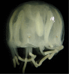
Tripedalia cystophora (a box jelly): this thumb-joint-sized jelly has four ‘sides’ and a cord-like structure on each side called a rhopalia. Each rhopalia has six eyes with two of those being camera style. Although the quality of discrimination cannot be good, this creature is a predator and uses visual cues to catch its prey.
Figure 7
Figure 7
Tripedalia cystophora (a box jelly): the terminal bulb of one of the rhopalia of this jelly showing a camera-style eye and the outline of a pit eye along the lower edge.
Figure 8
Figure 8
Histology of the eye of the nautilus: note the pinhole for focusing and the absence of the lens. Although the interpretation of the image would require much light, this mechanism would provide an image with surprising spatial information.
Figure 9
Figure 9
Histology of the lamprey eye (Petromyzon marinus). Although this is a basal vertebrate, the eye appears very much like a vertebrate eye. Note, however, the very thin cornea, which is separated from the overlying integument. Histologic section by Richard Dubielzig DVM.
Figure 10
Figure 10
Histology of the eye of a Bluefin tuna (Thunnus thynnus). Note that the equatorial diameter is ~60 mm and the anterior–posterior diameter measures ~40 mm although the eye has lost some size and volume during processing. This is a flat eye with a large round lens. Note also the ‘choroidal gland’ behind the macula, which is actually a plexus of blood vessels for nutrition and to provide warmth for the contents of the eye. Histologic section by Richard Dubielzig DVM.
Figure 11
Figure 11
Mopsus mormon (green jumping spider): note large anterior median eyes giving an appearance of a curious child. Some jumping spiders have tetrachromatic vision. The anterior lateral eyes can be seen on the creature, and these eyes have good vision as well.
Figure 12
Figure 12
Histology of jumping spider eye. Note the single lens in the ‘tube-like’ eye. The second refractive element in this eye is the convexiclivate fovea. The pit in the ‘retina’ causes light rays to diverge when striking the interface between the gel within the tube and ‘retina.’ Histologic section by Richard Dubielzig DVM.

References

    1. Reith M, Munholland J. Complete nucleotide sequence of the Porphyra purpurea chloroplast genome. Plant Mol Biol Rep 1995; 13: 333–335.
    1. McFadden GI. Chloroplast origin and integration. Plant Physiol 2001; 125: 50–53. - PMC - PubMed
    1. Feuda R, Hamilton SC, McInerney JO, Pisani D. Metazoan opsin evolution reveals a simple route to animal vision. Proc Natl Acad Sci USA 2012; 109: 18868–18872. - PMC - PubMed
    1. Grote M, Engelhard M, Hegemann P. Of ion pumps, sensors and channels-perspectives on microbial rhodopsins between science and history. Biochim Biophys Acta 2014; 1837: 533–545. - PubMed
    1. Porter ML. Beyond the eye: molecular evolution of extraocular photoreception. Integr Comp Biol 2016; 56: 842–852. - PubMed

LinkOut - more resources