Review
. 2018 Feb;32(2):287-301.
doi: 10.1038/eye.2017.222. Epub 2017 Oct 20.

Neuro-ophthalmic side effects of molecularly targeted cancer drugs

Affiliations
Review

Neuro-ophthalmic side effects of molecularly targeted cancer drugs

M T Bhatti et al. Eye (Lond). 2018 Feb.

Abstract

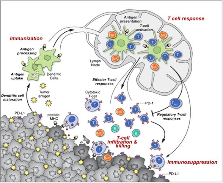
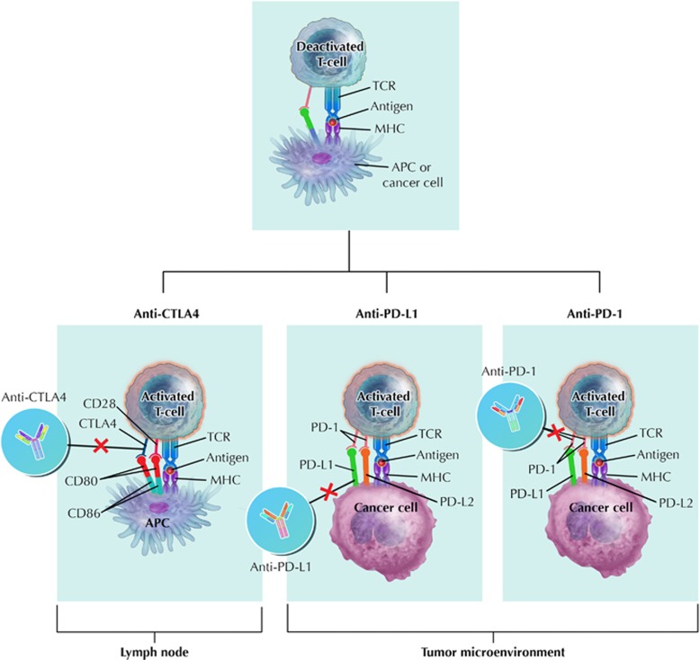
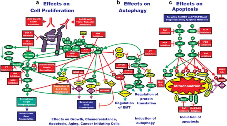
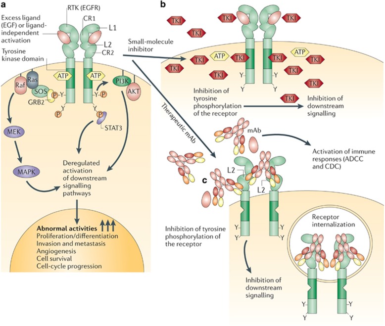
The past two decades has been an amazing time in the advancement of cancer treatment. Molecularly targeted therapy is a concept in which specific cellular molecules (overexpressed, mutationally activated, or selectively expressed proteins) are manipulated in an advantageous manner to decrease the transformation, proliferation, and/or survival of cancer cells. In addition, increased knowledge of the role of the immune system in carcinogenesis has led to the development of immune checkpoint inhibitors to restore and enhance cellular-mediated antitumor immunity. The United States Food and Drug Administration approval of the chimeric monoclonal antibody (mAb) rituximab in 1997 for the treatment of B cell non-Hodgkin lymphoma ushered in a new era of targeted therapy for cancer. A year later, trastuzumab, a humanized mAb, was approved for patients with breast cancer. In 2001, imatinib was the first small-molecule kinase inhibitor approved. The approval of ipilimumab-the first in class immune checkpoint inhibitor-in 2011 serves as a landmark period of time in the resurgence of immunotherapy for cancer. Despite the notion that increased tumor specificity results in decreased complications, toxicity remains a major hurdle in the development and implementation of many of the targeted anticancer drugs. This article will provide an overview of the current cellular and immunological understanding of cancer pathogenesis-the foundation upon which molecularly targeted therapies were developed-and a description of the ocular and neuro-ophthalmic toxicity profile of mAbs, immune checkpoint inhibitors, and small-molecule kinase inhibitors.


Conflict of interest statement

MTB: Novartis pharmaceuticals and Receptos.

AKSS: Bristol-Meyers Squibb, Celldex, Genentech, Merck, Immunocore, Reata.

Figures

Figure 1
Figure 1
Generation and regulation of antitumor immunity. Understanding the events in generating and regulating antitumor immunity suggests at least three sites for therapeutic intervention: promoting the antigen presentation functions of dendritic cells, promoting the production of protective T-cell responses and overcoming immunosuppression in the tumor bed. Antitumor immune responses must begin with the capture of tumor associated antigens by dendritic cells, either delivered exogenously or captured from dead or dying tumor cells. The dendritic cells process the captured antigen for presentation or cross-presentation on MHC class II and class I molecules, respectively, and migrate to draining lymph nodes. If capture and presentation occurred in the presence of an immunogenic maturation stimulus, dendritic cells will elicit anticancer effector T-cell responses in the lymph node; if no such stimulus was received, dendritic cells will instead induce tolerance leading to T-cell deletion, anergy or the production of Treg cells. In the lymph node, antigen presentation to T cells will elicit a response depending on the type of dendritic cell maturation stimulus received and on the interaction of T-cell co-stimulatory molecules with their surface receptors on dendritic cells. Thus, interaction of CD28 orOX40 with CD80/86 orOX40L will promote potentially protective T-cell responses, while interaction of CTLA4 with CD80/86 or PD-1 with PD-L1/PD-L2 will suppress T-cell responses, and possibly promote Treg formation. Antigen-educated T cells (along with B cells and NK cells) will exit the lymph node and enter the tumor bed, where a host of immunosuppressive defense mechanisms can be produced by tumors (or infiltrating myeloid cells) that oppose effector T-cell function. These include the upregulation of PD-L1/L2 on the cancer cell surface, release of PGE2, arginase and IDO (all T-cell suppressors), and the release of VEGF (triggered in part by intratumoral hypoxia), which inhibits T-cell diapedesis from the vasculature, and thus infiltration into the tumor bed. (Reprinted with permission: Mellman I, Coukos G, Dranoff G. Cancer immunotherapy comes of age. Nature. 2011 Dec 21; 480(7378): 480–489). A full color version of this figure is available at Eye online.
Figure 2
Figure 2
Site of immune checkpoint inhibitor action. Anti-CTLA4 prevents binding of CTLA4 to CD80 and CD86 ligands expressed on the surface of dendritic cells. The binding of CD28 to CD80 and CD86 ligands on the APC is a second co-stimulatory signal. CTLA-4 competes with CD28 in binding for CD80 and CD86 ligands. PD-L1 binds to PD-1 thereby de-activating T cells. Blocking either PD-L1 or PD-1 on cancer cells results in the activation of T cells. Anti-CTLA 4 action occurs in the lymph nodes therefore earlier on in the immune response, as compared to anti-PD-1, which is critical in the tumor microenvironment. APC, antigen presenting cell; CD, cluster of differentiation; MHC, major histocompatibility complex; PD, programmed death; PD-L, programmed death ligand; TCR, T-cell receptor. (Illustration by Rob Flewell, CMI).
Figure 3
Figure 3
Rationale for targeting both the Ras/Raf/MEK/ERK and Ras/PI3K/PTEN/Akt/mTOR pathways for suppressing cancer growth. (a) The Ras/Raf/MEK/ERK and Ras/PI3K/PTEN/Akt/mTOR pathways are both activated by upstream receptor ligation and frequently co-regulate many downstream targets in parallel. Thus for effective elimination of many cancers or prevention of aging, it may be necessary to target both signaling pathways. Activation of these pathways could also result in increased transcription of many genes that promote cellular growth and malignant transformation. (b) Inhibition of mTOR can result in the induction of autophagy, which is a very important mechanism of cell death, especially in solid tumors. (c) As described previously, both the Ras/Raf/MEK/ERK and Ras/PI3K/ PTEN/Akt/mTOR pathways regulate the activity of apoptotic proteins by post-translational mechanisms. Targeting this pathway may also contribute to the induction of apoptosis. Signaling molecules promoting phosphorylation events are indicated in green. Stimulatory signaling events are indicted in green lines with a green arrow before the target of the phophorylation. Small-molecule inhibitors are indicated in red. Inhibitory phosphorylation events are indicated in red lines with a block on the end before the target of the inhibition. Inhibitory signaling or proapoptotic molecules or inactivated molecules are indicated in yellow. A growth factor and a growth factor receptor are indicated in purple. Active transcription factors are indicated in purple diamonds. Inactivated transcription factors are indicated in yellow diamonds. (Reproduced with permission: Chappell WH, Steelman LS, Long JM, Kempf RC, Abrams SL, Franklin RA, Bäsecke J, Stivala F, Donia M, Fagone P, Malaponte G, Mazzarino MC, Nicoletti F, Libra M, Maksimovic-Ivanic D, Mijatovic S, Montalto G, Cervello M, Laidler P, Milella M, Tafuri A, Bonati A, Evangelisti C, Cocco L, Martelli AM, McCubrey JA. Ras/Raf/MEK/ERK and PI3K/PTEN/Akt/mTOR inhibitors: rationale and importance to inhibiting these pathways in human health. Oncotarget. 2011 Mar; 2(3): 135–164). A full color version of this figure is available at Eye online.
Figure 4
Figure 4
Distinct mechanisms of small-molecule inhibitors and monoclonal antibodies for targeting receptor tyrosine kinases in cancer cells. (a). Epidermal growth factor receptor (EGFR) and receptor tyrosine kinase (RTK)-dependent growth signaling in cancer cells. The extracellular region of EGFR consists of four domains, the ligand-binding domains (L1 and L2) and the cysteine-rich domains (CR1 and CR2), and the C-terminal domain of EGFR contains six tyrosine residues (Y; only two are depicted here for simplicity). Following the activation of EGFR by ligand binding or ligand-independent dimerization, the Ras–Raf–MEK–MAPK pathway is activated through the growth factor receptor bound protein 2 (GRB2)–SOS complex. EGFR-mediated signaling also activates the phosphatidylinositol 3-kinase (PI3K)– AKT pathway, which contributes to anti-apoptotic effects of EGFR activation. In addition, signal transducer and activator of transcription (Stat) proteins (STAT1, STAT3, and STAT5) are also activated. The coordinated effects of these EGFR downstream signaling pathways lead to the induction of cellular responses including proliferation, differentiation, cell motility, adhesion, and angiogenesis. The deregulation of EGFR-mediated signaling in some cancer cells leads to aberrant proliferation, invasion, metastasis, and neovascularization. (b) Small-molecule tyrosine kinase inhibitors (TKIs) such as gefitinib function as ATP analogues and inhibit EGFR signaling by competing with ATP binding within the catalytic kinase domain of RTKs. As a result, the activation of various downstream signaling pathways is blocked. Each TKI has a different selectivity for RTKs, and some are dual- or multi-selective, which might provide a therapeutic advantage. (c) By contrast, therapeutic monoclonal antibodies (mAbs) bind to the ectodomain of the RTK with high specificity (for example, cetuximab binds to the L2 domain of EGFR, and thereby inhibits its downstream signaling by triggering receptor internalization and hindering ligand–receptor interaction. Unlike small-molecule inhibitors, mAbs also activate Fcγ- receptor-dependent phagocytosis or cytolysis by immune-effector cells such as neutrophils, macrophages and natural killer cells by inducing complement-dependent cytotoxicity (CDC) or antibody-dependent cellular cytotoxicity (ADCC). MAPK, mitogen-activated protein kinase; MEK, mitogen-activated protein kinase kinase. (Reproduced with permission: Imai K, Takaoka A. Comparing antibody and small-molecule therapies for cancer. Nat Rev Cancer. 2006 Sep; 6(9): 714–727). A full color version of this figure is available at Eye online.

References

    1. Siegel RL, Miller KD, Jemal A. Cancer Statistics, 2017. CA Cancer J Clin 2017; 67(1): 7–30. - PubMed
    1. Gerber DE. Targeted therapies: a new generation of cancer treatments. Am Fam Physician 2008; 77(3): 311–319. - PubMed
    1. Aggarwal S. Targeted cancer therapies. Nat Rev Drug Discov 2010; 9(6): 427–428. - PubMed
    1. Kocher R, Roberts B. The calculus of cures. N Engl J Med 2014; 370(16): 1473–1475. - PubMed
    1. Scharf O, Colevas AD. Adverse event reporting in publications compared with sponsor database for cancer clinical trials. J Clin Oncol 2006; 24(24): 3933–3938. - PubMed

MeSH terms

Substances