. 2018 Feb 15;197(4):470-480.
doi: 10.1164/rccm.201706-1161OC.

Red Blood Cells Homeostatically Bind Mitochondrial DNA through TLR9 to Maintain Quiescence and to Prevent Lung Injury

Affiliations

Red Blood Cells Homeostatically Bind Mitochondrial DNA through TLR9 to Maintain Quiescence and to Prevent Lung Injury

Meghan J Hotz et al. Am J Respir Crit Care Med. .

Abstract

Rationale: Potentially hazardous CpG-containing cell-free mitochondrial DNA (cf-mtDNA) is routinely released into the circulation and is associated with morbidity and mortality in critically ill patients. How the body avoids inappropriate innate immune activation by cf-mtDNA remains unknown. Because red blood cells (RBCs) modulate innate immune responses by scavenging chemokines, we hypothesized that RBCs may attenuate CpG-induced lung inflammation through direct scavenging of CpG-containing DNA.

Objectives: To determine the mechanisms of CpG-DNA binding to RBCs and the effects of RBC-mediated DNA scavenging on lung inflammation.

Methods: mtDNA on murine RBCs was measured under basal conditions and after systemic inflammation. mtDNA content on human RBCs from healthy control subjects and trauma patients was measured. Toll-like receptor 9 (TLR9) expression on RBCs and TLR9-dependent binding of CpG-DNA to RBCs were determined. A murine model of RBC transfusion after CpG-DNA-induced lung injury was used to investigate the role of RBC-mediated DNA scavenging in mitigating lung injury in vivo.

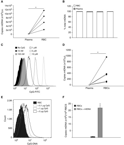
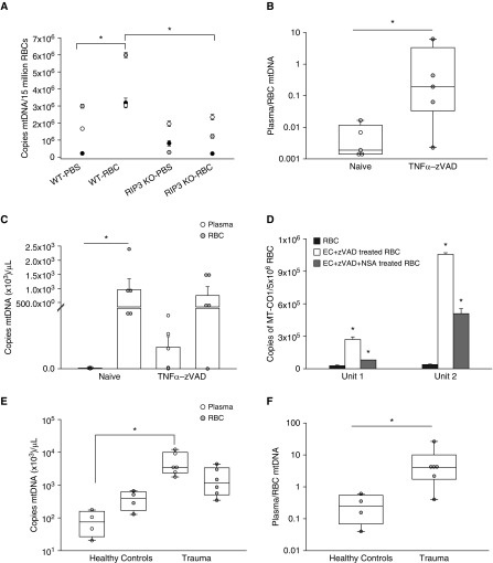
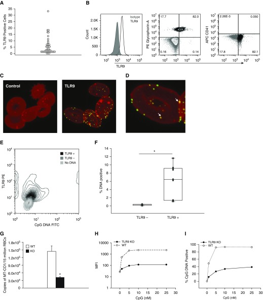
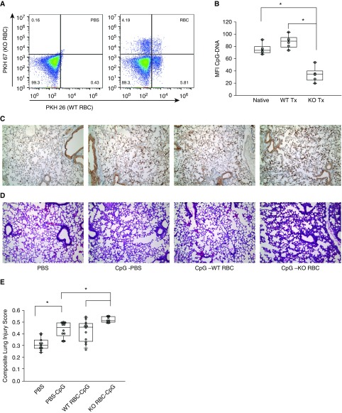
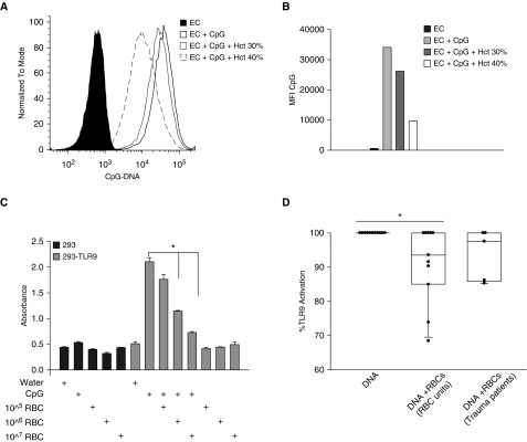
Measurements and main results: Under basal conditions, RBCs bind CpG-DNA. The plasma-to-RBC mtDNA ratio is low in naive mice and in healthy volunteers but increases after systemic inflammation, demonstrating that the majority of cf-mtDNA is RBC-bound under homeostatic conditions and that the unbound fraction increases during inflammation. RBCs express TLR9 and bind CpG-DNA through TLR9. Loss of TLR9-dependent RBC-mediated CpG-DNA scavenging increased lung injury in vivo.

Conclusions: RBCs homeostatically bind mtDNA, and RBC-mediated DNA scavenging is essential in mitigating lung injury after CpG-DNA. Our data suggest a role for RBCs in regulating lung inflammation during disease states where cf-mtDNA is elevated, such as sepsis and trauma.

Keywords: CpG-DNA; RBC; Toll-like receptor 9; mitochondrial DNA.


Figures

Figure 1.
Figure 1.
Mitochondrial DNA (mtDNA) is present on human and murine red blood cells (RBCs) under basal conditions and partitions with RBCs rather than the soluble fraction. (A) Cell-free mtDNA partitions with RBCs rather than plasma in healthy human subjects: *P = 0.029, n = 4. (B) Percentage of total mtDNA is greater in RBCs than in plasma for each healthy subject tested (98.67 vs. 1.30% for RBCs vs. plasma; P < 0.001). (C) Human RBCs bind CpG-DNA under basal conditions. (D) Cell-free mtDNA partitions with RBCs rather than plasma in naive mice: *P = 0.008, n = 5 mice, two independent studies. (E) Murine RBCs bind CpG-DNA. (F) Murine RBCs were incubated with 200 pg of purified murine Mt-Co1 (gene encoding cytochrome c oxidase subunit 1) amplicon for 1 hour at 37°C. The cells were then washed, lysates were prepared, and PCR for Mt-Co1 was performed. Murine RBCs acquired mtDNA: *P < 0.01. Bar graph shows mean ± SEM. The Mann-Whitney rank sum test was used where data were not normally distributed. FITC = fluorescein isothiocyanate.
Figure 2.
Figure 2.
Mitochondrial DNA (mtDNA) accumulates on human and murine red blood cells (RBCs) after necroptosis, and the plasma-to-RBC mtDNA ratio is elevated after systemic inflammation. (A) Murine RBCs bind mtDNA after necroptosis: mtDNA on RBCs obtained from phosphate-buffered saline (PBS)–treated versus RBC-transfused mice. mtDNA is increased on RBCs obtained from transfused mice. mtDNA is higher on RBCs obtained from wild-type (WT) transfused mice when compared with receptor-interacting serine/threonine protein kinase 3 knockout (RIP3 KO) transfused mice. Each symbol represents three mice and each shade represents an individual study: n = 9 mice per group, three independent studies, *P = 0.05. (B) The plasma-to-RBC mtDNA ratio is increased 2 hours after systemic inflammation. mtDNA in naive and TNFα–zVAD–treated mice: Mann-Whitney rank sum test, *P = 0.032. (C) There is increased mtDNA in the RBC fraction when compared with the plasma of naive mice (*P = 0.008) but not after systemic inflammation: n = 5 mice, two independent studies for B and C. (D) Human RBCs bind mtDNA released after lung endothelial cell necroptosis. Preincubation with the necroptosis inhibitor necrosulfonamide (NSA) attenuates mtDNA acquisition by RBCs: *P < 0.01. PCR results for MT-CO1 are shown. Three units were tested, and two representative units are shown (Unit 1 and Unit 2). (E) RBC and plasma mtDNA in healthy subjects and trauma patients: n = 4 healthy subjects, n = 6 trauma patients, *P = 0.0381. (F) The plasma-to-RBC mtDNA ratio is increased after trauma or systemic inflammation: *P = 0.019, Mann-Whitney rank sum test. Bar graphs show mean ± SEM. Box-and-whisker plots show median, interquartile range, and maximum and minimum values. EC = endothelial cells; MT-CO1 = mitochondrially encoded cytochrome c oxidase 1; TNF = tumor necrosis factor; zVAD = {benzyloxycarbonyl-l-valyl-l-alanyl-[(2S)-2-amino-3-(methoxycarbonyl)propionyl]}fluoromethane.
Figure 3.
Figure 3.
Human and murine red blood cells (RBCs) bind CpG-DNA through Toll-like receptor 9 (TLR9). (A) TLR9 expression on RBCs obtained from healthy human donors and leukoreduced RBC units: n = 39. (B) TLR9-positive cells obtained from a healthy donor are CD235 positive and CD41 negative. (C) Confocal imaging confirms the presence of TLR9 on human RBCs: green, TLR9; red, membrane stain (PKH); secondaries alone are shown on the left. (D) Arrows depict areas of colocalization of TLR9 with RBC membrane. (E and F) TLR9-positive RBCs bind more CpG-DNA than TLR9-negative RBCs: *P < 0.001, Mann-Whitney rank sum test. RBCs not treated with DNA are also depicted in E; nine individual RBC preparations from healthy donors or RBC units were tested. (G) RBCs obtained from naive wild-type (WT) mice contain more mitochondrial DNA than do RBCs obtained from naive TLR9 knockout (KO) mice: *P = 0.053, n = 5 or 6 mice per group, one study of three shown. (H and I) WT and TLR9 KO RBCs were labeled with PKH26 and PKH67 and mixed with CpG-DNA. TLR9 KO RBCs (solid line and solid circles) bind CpG-DNA less efficiently than WT RBCs (dashed line and open circles). (H) MFI versus CpG: 0–25 nM CpG doses shown. (I) Percent CpG-DNA–positive cells versus CpG concentration for WT and TLR9 KO RBCs. Bar graph shows mean ± SEM. Box-and-whisker plot shows median, interquartile range, and maximum and minimum values. APC = allophycocyanin; FITC = fluorescein isothiocyanate; MFI = mean fluorescence intensity; MT-CO1 = mitochondrially encoded cytochrome c oxidase I; PE = phycoerythrin; PKH = membrane dye.
Figure 4.
Figure 4.
Red blood cells (RBCs) scavenge CpG-DNA in vivo, and loss of RBC Toll-like receptor 9 (TLR9)–mediated CpG-DNA scavenging augments lung injury. (A) Left: Spleen RBCs obtained from mice treated with phosphate-buffered saline (PBS) and CpG-DNA. Right: PKH-positive cells are detected in the RBC–CpG-DNA–treated animals. (B) Wild-type (WT) transfused RBCs have increased CpG positivity when compared with native RBCs in the spleen (*P = 0.043); knock-out (KO) RBCs are less CpG positive when compared with native and transfused WT RBCs (*P < 0.001, n = 6 mice per group, two independent studies). (CE) Loss of RBC-mediated CpG-DNA scavenging augments lung injury. Lungs obtained from mice treated with PBS, CpG-DNA, CpG-DNA + WT RBCs, and CpG-DNA + TLR9-KO RBCs were compared. WT or KO RBC transfusions (10 μl/g; hematocrit, 65%) were given simultaneously with CpG-DNA. (C) Ly6G; (D) hematoxylin and eosin; and (E) composite lung injury scores demonstrate increased tissue injury in CpG TLR9-KO RBC-treated mice when compared with all other groups: *P < 0.001, n = 7–12 mice per group, three independent studies. Box-and-whisker plots show median, interquartile range, and maximum and minimum values. MFI = mean fluorescence intensity; PKH = membrane dye; Tx = transfused.
Figure 5.
Figure 5.
Red blood cells (RBCs) attenuate CpG-induced Toll-like receptor 9 (TLR9) activation in vitro. (A) RBCs scavenge CpG-DNA (100 nM) from human lung microvascular endothelial cells. FITC–CpG binding to endothelial cells (ECs) or to ECs treated with CpG-DNA in the presence or absence of RBCs is shown. EC, EC + CpG-DNA, EC + CpG-DNA + RBCs (Hct, 30 or 40% is shown). (B) MFI of CpG-DNA in the treatments in A: n = 3 independent studies, two of the three RBC units scavenged CpG-DNA, and one representative study is shown. (C) CpG-DNA–induced activation of TLR9 was assayed with 293 cells transfected with a TLR9 reporter. CpG-DNA (10 μg/ml) induced activation of TLR9, which was attenuated with the addition of RBCs; *P < 0.001. (D) RBCs from RBC units attenuated CpG-DNA–induced TLR9 activation, whereas RBCs obtained from trauma patients do not attenuate TLR9 activation: 11 units tested, *P = 0.048; n = 5 trauma patients, P = 0.160. Bar graph shows mean ± SEM. Box-and-whisker plot shows median, interquartile range, and maximum and minimum values. FITC = fluorescein isothiocyanate; Hct = hematocrit; MFI = mean fluorescence intensity.

Comment in

Similar articles

Cited by

References

    1. Stroun M, Lyautey J, Lederrey C, Olson-Sand A, Anker P. About the possible origin and mechanism of circulating DNA apoptosis and active DNA release. Clin Chim Acta. 2001;313:139–142. - PubMed
    1. Snyder MW, Kircher M, Hill AJ, Daza RM, Shendure J. Cell-free DNA comprises an in vivo nucleosome footprint that informs its tissues-of-origin. Cell. 2016;164:57–68. - PMC - PubMed
    1. Nakahira K, Kyung SY, Rogers AJ, Gazourian L, Youn S, Massaro AF, et al. Circulating mitochondrial DNA in patients in the ICU as a marker of mortality: derivation and validation. PLoS Med. 2013;10:e1001577. - PMC - PubMed
    1. Simmons JD, Lee YL, Mulekar S, Kuck JL, Brevard SB, Gonzalez RP, et al. Elevated levels of plasma mitochondrial DNA DAMPs are linked to clinical outcome in severely injured human subjects. Ann Surg. 2013;258:591–596, discussion 596–598. - PMC - PubMed
    1. Steinman CR. Circulating DNA in systemic lupus erythematosus: isolation and characterization. J Clin Invest. 1984;73:832–841. - PMC - PubMed

Publication types

MeSH terms