. 2017 Oct 1;58(12):5399-5411.
doi: 10.1167/iovs.17-22978.

Concepts and Strategies in Retinal Gene Therapy

Affiliations

Concepts and Strategies in Retinal Gene Therapy

Gustavo D Aguirre. Invest Ophthalmol Vis Sci. .
No abstract available


Figures

Figure 1
Figure 1
Discovery approaches for canine retinal degeneration loci and genes. The cumulative numbers of loci and causative mutations identified are shown, classified according to the discovery approach used. Note that the chromosomal location and the mutation were reported concurrently for cd, cord1, osd1, and osd2. *New mutation in a gene for which a mutation is already known. Figure courtesy of Keiko Miyadera.
Figure 2
Figure 2
Targeting photoreceptors in the CNGB3−/− mutant (A1–A3) and normal (B1–B3) dog retina, and in normal nonhuman primate (NHP, M. fascicularis; C1–C3) using AAV5 vectors with different promoters. (A1) Four weeks after subretinal injection using PR2.1 promoter, there is robust native GFP expression (green) in L/M-cones (red), which is the predominant cone class in the canine retina (scale bar: 40 μm). Figure from Komaromy AM, Alexander JJ, Rowlan JS, et al. Gene therapy rescues cone function in congenital achromatopsia. Hum Mol Genet. 2010;19:2581–2593. © The Author 2010. Reprinted with permission from Oxford University Press. (A2, A3) The hybrid GNAT2/IRBP promoter results in robust GFP expression in both L/M- (A2, red) and S- (A3, red) cones (Figs. A2, A3 courtesy of András Komaromy). (B1–B3) The hIRBP promoter targets native GFP expression (green) after injection of a 1.5 × 10¹¹ μg/mL titer. GFP expression is low after 2 weeks (B1), and increases by 8 weeks post injection (B2); at 8 weeks, hCAR labeling (red) confirms expression in cones (B3). Scale bar: 20 μm. Figures B1–B3 reprinted from Beltran WA, Cideciyan AV, Lewin AS, et al. Gene therapy rescues photoreceptor blindness in dogs and paves the way for treating human X-linked retinitis pigmentosa. Proc Natl Acad Sci U S A. 2012;109:2132–2137. © 2012 The Authors. (C1–C3) Peripheral retina from NHP after subretinal injection using hRGK1 promoter to target eGFP (green) expression to rods and cones. hCAR antibody labels cones (red; C1), and colocalizes with eGFP in cones (C3). White arrows identify the same cone cells in the three images; scale bar: 17 μm. ONL, outer nuclear layer. Figures C1–C3 reprinted with permission from Beltran WA, Cideciyan AV, Boye SE, et al. Optimization of retinal gene therapy for X-linked retinitis pigmentosa due to RPGR mutations. Mol Ther. 2017;25:1866–1880. © 2017 The American Society of Gene and Cell Therapy.
Figure 3
Figure 3
I. Long-term efficacy of rescued rod and cone retinal function in RPE65 mutants treated at an early disease stage and evaluated up to a decade later (A–D), or treated late and evaluated 1 month after the treatment (E). (A) Dark-adapted (DA) ERGs evoked by standard white flashes (black traces), or under light-adapted (LA) (red traces) conditions. Black vertical lines show the timing of the flashes. (B) ERG photoresponses evoked by white flashes of high energy under dark-adapted conditions; same data are shown on slow (upper) and fast (lower) time scales. Gray lines show the baseline and the 4-ms time point where rod photoreceptor responses were measured. (C) Comparison of rod and cone function in the treated RPE65-mutant dogs (Tx; green triangles) compared with previously published normal (N) and untreated control (Ctrl) eyes. Rod function shown refers to the DA ERG photoresponse amplitude at 4 ms, and cone function refers to the peak amplitude of the LA 29-Hz waveform. Horizontal dashed lines represent the upper limit (mean + 3 SD) of the respective measurement in the group of untreated control eyes. (D) Durability of the rod and cone ERG amplitudes in dog D23 treated a decade earlier. Subretinally treated right eye is shown with green symbols, and the intravitreally treated left eye is shown with gray symbols. Lines show previously published data extending to age 3 years. (E) Bilateral ERGs recorded with similar methods and stimuli as shown in (A) in a normal dog and three unilaterally treated older RPE65-mutant dogs (age range from 4.9 to 6.6 years). ERG recordings performed 1 month later show definite treatment effects in the eyes with gene therapy. II. Gene therapy outcomes in RPE65-mutant dogs treated before (A1–A5) and after (B1–B5) the onset of retinal degeneration. (A1, A2) Photoreceptor (ONL) thickness topography in two dogs treated before the onset of the degeneration and evaluated ∼5 to 9 years later. There is retention of ONL thickness within the treatment region (dashed lines). (A3–A5) ONL thickness quantified as a function of age at five retinal locations in five dogs treated before the initiation of degeneration. Red symbols correspond to retinal locations outside the treatment region, and green symbols correspond to locations within the treatment region. (B1, B2) ONL thickness topography in two dogs treated after the onset of degeneration. There is no evidence for thicker ONL within the treatment regions compared with outside the treatment regions. (B3–B5) ONL thickness quantified as a function of age at five retinal locations in treated eyes. Both untreated control (red symbols) and treated (green symbols) regions are not substantially different compared with the natural history of disease. III. Retina of dog D23 treated at 0.3 year before the onset of retinal degeneration shows remarkable rescue of photoreceptors from degeneration when assessed over a decade later. (A) Schematic representation of the en face image showing the treatment area (dashed lines), on which is superimposed the ONL rows (mean of three values in each area sampled) and disease staging (a, advanced atrophy with gliosis and loss of retinal layer organization; m, moderate photoreceptor loss with 1/3 to 1/2 of ONL remaining; n, normal) assessed at 11.2 years. (B–D) Representative images taken from areas identified in (A). In the treatment area, there is normal retinal preservation (B), although the RPE shows vacuolated inclusions typical of the disease (arrowheads). At the edge of the treatment border (C), the photoreceptor layer becomes markedly attenuated and is absent (D) outside of the treatment region. Double immunolabeling with RPE65 (red) and rod opsin (green) taken from region corresponding to (B). RPE labeling: RPE is present inside the treatment region, and rod outer segment labeling is distinct. RPE, retinal pigment epithelium; GCL, ganglion cell layer; NFL, nerve fiber layer; OPL, outer plexiform layer; OS/IS, outer and inner segment layer; INL, inner nuclear layer; IPL, inner plexiform layer; scale bar: 20 μm. Figures and legends modified from Cideciyan AV, Jacobson SG, Beltran WA, et al. Human retinal gene therapy for Leber congenital amaurosis shows advancing retinal degeneration despite enduring visual improvement. Proc Natl Acad Sci U S A. 2013;110:E517–E525. © 2013 The Authors.
Figure 4
Figure 4
Gene therapy outcomes in CNGB3-achromatopsia. (A) CNGB3 mutants with either a missense mutation (m/m) or genomic deletion (−/−) show normal rod ERG responses, but absent cone responses. Gene therapy restores the cone ERG responses (far right column), and the effect is sustained for at least 2.5 years (B). (C) Cone ERG flicker amplitude increased with higher hCNGB3 transgene expression. Dogs with no recovery of cone function had low levels of transgene expression and were treated with the less robust 3LCR-PR0.5 promoter (red circle, treatment age 8, 23, 28 weeks; green circle, treatment age 60–81 weeks). The optimal PR2.1 promoter resulted in high levels of transgene expression in one dog (blue circle), but no cone function rescue when treatment was done at 54 weeks. Figures 4A–C reprinted from Komaromy AM, Alexander JJ, Rowlan JS, et al. Gene therapy rescues cone function in congenital achromatopsia. Hum Mol Genet. 2010;19:2581–2593. © 2010 The Author. Reprinted with permission from Oxford University Press. (D) Photoreceptor deconstruction with CNTF. The relative amounts of retinal hCNGB3 mRNA expression were comparable and not significantly different when subretinal AAV injections were preceded by either intravitreal PBS (no cone function recovery) or CNTF (cone function recovery). (E) In the wild-type retina, CNGA3 and GNAT2 colocalize with L/M opsin in the cone outer segment (top). Gene therapy following intravitreal PBS (middle) fails to correct the mislocalization of CNGA3 and GNAT2 from the outer segment (middle). However, pretreatment with CNTF 1 week prior to gene therapy corrects the mislocalization in the now functional L/M cones (middle). Scale bar: 10 μm. Figures 4D, 4E reprinted with permission from Komaromy AM, Rowlan JS, Corr AT, et al. Transient photoreceptor deconstruction by CNTF enhances rAAV-mediated cone functional rescue in late stage CNGB3-achromatopsia. Mol Ther. 2013;21:1131–1141. © 2013 The American Society of Gene & Cell Therapy.
Figure 5
Figure 5
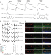
I. Retinal disease phenotypes caused by RPGR-ORF15 mutations in human patients and in dogs. (A) Different patterns of photoreceptor topography in two XLRP patients with RPGR mutations. ONL thickness topography is mapped to a pseudocolor scale. (Inset) Representative normal subject. Locations of fovea and optic nerve (ON) are shown. (B) Different patterns of photoreceptor topography in the canine models of RPGR-ORF15; mapping as performed with the human data. (Inset) Map of a representative wild-type dog with location of ON labeled. (C) ONL thickness profile along the vertical meridian (Inset) comparing XLPRA1 and XLPRA2 of different ages (thin traces) versus normal results (gray band). Mean (±SD) results are from groups of younger (7–28 weeks) and older (36–76 weeks) dogs. The thicker red line represents the data from the oldest dogs examined (>144 weeks old). Brackets mark the location of the high photoreceptor density corresponding to the canine visual streak. Figures and legends in I modified from Beltran WA, Cideciyan AV, Lewin AS, et al. Gene therapy rescues photoreceptor blindness in dogs and paves the way for treating human X-linked retinitis pigmentosa. Proc Natl Acad Sci U S A. 2012;109:2132–2137. © 2012 The Authors. II, III. Efficacy and long-term stability of gene therapy intervention at (II) mid-stage and (III) late-stage disease. (A) Pseudocolor maps of ONL thickness topography in XLPRA2 dogs treated at 12 (mid-stage) and 26 (late-stage) weeks of age. Dashed outline is the retinal region corresponding to the subretinal vector bleb at treatment. Schematic, right, paired loci across the treatment boundary and in the inferior retina chosen for quantitative evaluation. Eyes are shown as equivalent right eyes with optic nerve and major blood vessels overlaid for ease of comparability. T, temporal; N, nasal retina. (B) Progressive changes in ONL fraction recorded serially between 11 (mid-stage) and 25 (late-stage) weeks through to 130 weeks of age in treated (green) and untreated (red) loci in the superior and inferior retinas of three XLPRA2 dogs treated for each disease stage. None of the three late-stage treated eyes received injection in the inferior retina; thus, only untreated loci are shown in inferior retina. Vertical green arrows depict the timing of treatment. Dashed lines show the range of ONL fraction expected in wild-type eyes or natural history of progression in untreated XLPRA2 eyes. Smaller symbols represent the individual data and larger symbols with error bars represent mean ± SD; *P < 0.01 for paired t-tests between treated and untreated loci. (C) Retinal morphology at 113 weeks of age in the untreated (UnTx) and treated (Tx) areas of a dog injected at mid- and late-stage disease and immunohistochemistry labeling of stable human RPGR transgene product, which is present only in treated areas. IV. Long-term durability of retinal function after gene therapy intervention at late-stage disease. Representative ERG traces of rod and mixed rod–cone responses recorded dark-adapted and cone responses to single stimuli, or 29-Hz cone flicker recorded light-adapted. Figures and legends in II, III, and IV modified from Beltran WA, Cideciyan AV, Iwabe S, et al. Successful arrest of photoreceptor and vision loss expands the therapeutic window of retinal gene therapy to later stages of disease. Proc Natl Acad Sci U S A. 2015;112:E5844–E5853. © 2015 The Authors.

References

    1. Bramall AN, Wright AF, Jacobson SG, McInnes RR. . The genomic, biochemical, and cellular responses of the retina in inherited photoreceptor degenerations and prospects for the treatment of these disorders. Annu Rev Neurosci. 2010; 33: 441– 472. - PubMed
    1. Wright AF, Chakarova CF, Abd El-Aziz MM, Bhattacharya SS. . Photoreceptor degeneration: genetic and mechanistic dissection of a complex trait. Nat Rev Genet. 2010; 11: 273– 284. - PubMed
    1. Acland GM, Aguirre GD, Ray J,et al. . Gene therapy restores vision in a canine model of childhood blindness. Nat Genet. 2001; 28: 92– 95. - PubMed
    1. Acland GM, Aguirre GD, Bennett J,et al. . Long-term restoration of rod and cone vision by single dose rAAV-mediated gene transfer to the retina in a canine model of childhood blindness. Mol Ther. 2005; 12: 1072– 1082. - PMC - PubMed
    1. Cideciyan AV, Jacobson SG, Beltran WA,et al. . Human retinal gene therapy for Leber congenital amaurosis shows advancing retinal degeneration despite enduring visual improvement. Proc Natl Acad Sci U S A. 2013; 110: E517– E525. - PMC - PubMed

Publication types