

Less is more: neural mechanisms underlying anomia treatment in chronic aphasic patients
- PMID: 29053773
- PMCID: PMC5808641
- DOI: 10.1093/brain/awx234
Less is more: neural mechanisms underlying anomia treatment in chronic aphasic patients
Abstract
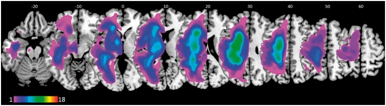
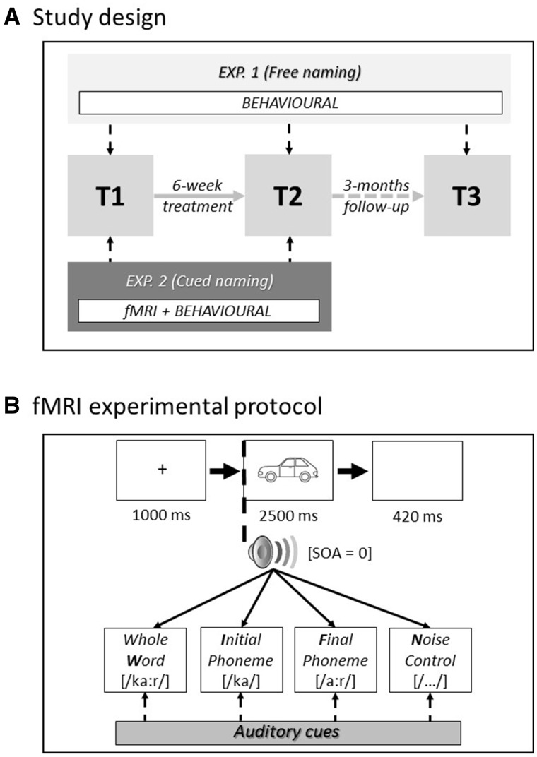
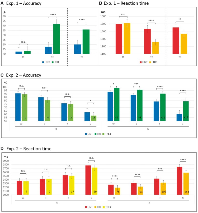
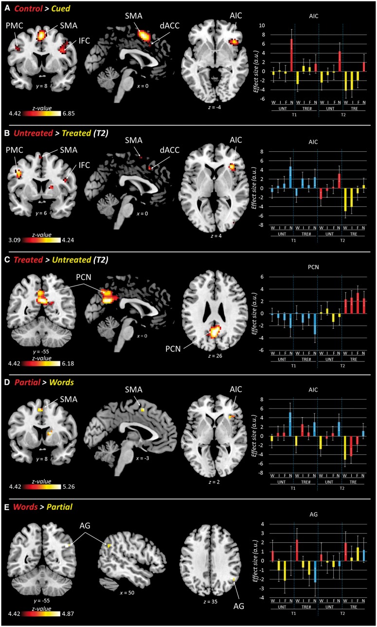
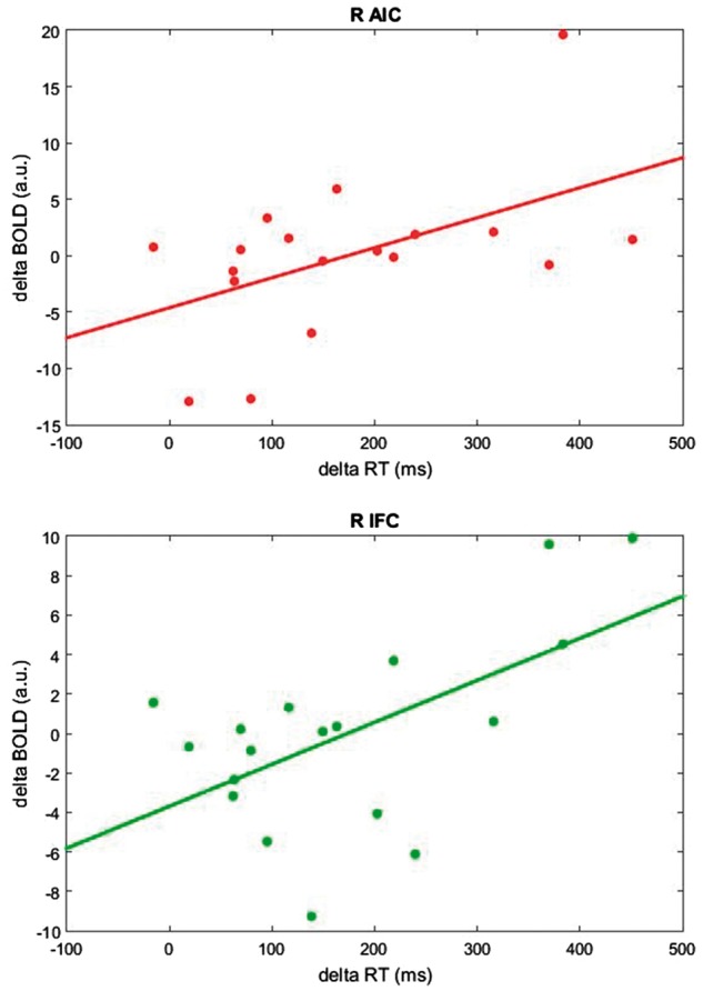
See Thompson and Woollams (doi:10.1093/brain/awx264) for a scientific commentary on this article. Previous research with aphasic patients has shown that picture naming can be facilitated by concurrent phonemic cueing [e.g. initial phoneme(s) of the word that the patient is trying to retrieve], both as an immediate word retrieval technique, and when practiced repeatedly over time as a long-term anomia treatment. Here, to investigate the neural mechanisms supporting word retrieval, we adopted—for the first time—a functional magnetic resonance imaging task using the same naming procedure as it occurs during the anomia treatment process. Before and directly after a 6-week anomia treatment programme, 18 chronic aphasic stroke patients completed our functional magnetic resonance imaging protocol—a picture naming task aided by three different types of phonemic cues (whole words, initial phonemes, final phonemes) and a noise-control condition. Patients completed a naming task based on the training materials, and a more general comprehensive battery of language tests both before and after the anomia treatment, to determine the effectiveness and specificity of the therapy. Our results demonstrate that the anomia treatment was effective and specific to speech production, significantly improving both patients’ naming accuracy and reaction time immediately post-treatment (unstandardized effect size: 29% and 17%, respectively; Cohen’s d: 3.45 and 1.83). Longer term gains in naming were maintained 3 months later. Functional imaging results showed that both immediate and long-term facilitation of naming involved a largely overlapping bilateral frontal network including the right anterior insula, inferior frontal and dorsal anterior cingulate cortices, and the left premotor cortex. These areas were associated with a neural priming effect (i.e. reduced blood oxygen level-dependent signal) during both immediate (phonemically-cued versus control-cue conditions), and long-term facilitation of naming (i.e. treated versus untreated items). Of note is that different brain regions were sensitive to different phonemic cue types. Processing of whole word cues was associated with increased activity in the right angular gyrus; whereas partial word cues (initial and final phonemes) recruited the left supplementary motor area, and right anterior insula, inferior frontal cortex, and basal ganglia. The recruitment of multiple and bilateral areas may help explain why phonemic cueing is such a successful behavioural facilitation tool for anomia treatment. Our results have important implications for optimizing current anomia treatment approaches, developing new treatments, and improving speech outcome for aphasic patients.
Keywords: anomia treatment; aphasia; phonemic cueing; picture naming; word retrieval.
Figures
Comment in
-
Reduced neural 'effort' after naming treatment in anomia.Brain. 2017 Nov 1;140(11):2773-2775. doi: 10.1093/brain/awx264. Brain. 2017. PMID: 29088349 No abstract available.
References
-
- Abel S, Weiller C, Huber W, Willmes K. Neural underpinnings for model-oriented therapy of aphasic word production. Neuropsychologia 2014; 57: 154–65. - PubMed
-
- Abel S, Weiller C, Huber W, Willmes K, Specht K. Therapy-induced brain reorganization patterns in aphasia. Brain 2015; 138: 1097–112. - PubMed
-
- Bamiou DE, Musiek FE, Luxona LM. The insula (Island of Reil) and its role in auditory processing: literature review. Brain Res Brain Res Rev 2003; 42: 143–54. - PubMed
-
- Best W, Greenwood A, Grassly J, Herbert R, Hickin J, Howard D. Aphasia rehabilitation: does generalisation from anomia therapy occur and is it predictable? A case series study. Cortex 2013; 49: 2345–57. - PubMed
Publication types
MeSH terms
Grants and funding
LinkOut - more resources
Full Text Sources
Other Literature Sources
Medical
