Comparative Study
. 2017 Dec 15;292(50):20669-20682.
doi: 10.1074/jbc.M117.792911. Epub 2017 Oct 20.

The type II transmembrane serine protease matriptase cleaves the amyloid precursor protein and reduces its processing to β-amyloid peptide

Affiliations
Comparative Study

The type II transmembrane serine protease matriptase cleaves the amyloid precursor protein and reduces its processing to β-amyloid peptide

Erwan Lanchec et al. J Biol Chem. .

Abstract

Recent studies have reported that many proteases, besides the canonical α-, β-, and γ-secretases, cleave the amyloid precursor protein (APP) and modulate β-amyloid (Aβ) peptide production. Moreover, specific APP isoforms contain Kunitz protease-inhibitory domains, which regulate the proteolytic activity of serine proteases. This prompted us to investigate the role of matriptase, a member of the type II transmembrane serine protease family, in APP processing. Using quantitative RT-PCR, we detected matriptase mRNA in several regions of the human brain with an enrichment in neurons. RNA sequencing data of human dorsolateral prefrontal cortex revealed relatively high levels of matriptase RNA in young individuals, whereas lower levels were detected in older individuals. We further demonstrate that matriptase and APP directly interact with each other and that matriptase cleaves APP at a specific arginine residue (Arg-102) both in vitro and in cells. Site-directed (Arg-to-Ala) mutagenesis of this cleavage site abolished matriptase-mediated APP processing. Moreover, we observed that a soluble, shed matriptase form cleaves endogenous APP in SH-SY5Y cells and that this cleavage significantly reduces APP processing to Aβ40. In summary, this study identifies matriptase as an APP-cleaving enzyme, an activity that could have important consequences for the abundance of Aβ and in Alzheimer's disease pathology.

Keywords: Alzheimer disease; ST14; amyloid precursor protein (APP); amyloid-β (Aβ); brain; enzyme processing; extracellular matrix protein; matriptase; serine protease.


Conflict of interest statement

The authors declare that they have no conflicts of interest with the contents of this article

Figures
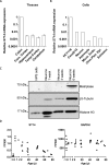
Figure 1.
Figure 1.
Matriptase (ST14) mRNA relative expression in human brains and nervous system cells. A, levels of ST14 mRNA were analyzed in the human frontal (Fron.) cortex (n = 18), temporal (Temp.) cortex (n = 8), hippocampus (n = 5), and cerebellum (n = 7) and expressed relative to that in the human colon tissue (n = 3). The difference between the different brain regions was not significant (Student's t test, p > 0.05). Error bars represent means ± S.D. B, the levels of ST14 mRNA were analyzed in human neurons, astrocytes, microvascular endothelial cells (Micro. Endo.), choroid plexus epithelial (Chor. Plex. Epi.) cells, Schwann cells, and epithelial colorectal adenocarcinoma Caco-2/15 cells and expressed relative to human colon carcinoma HCT116 cells (triplicate analyses were performed on each sample). C, expression of matriptase (immunoblot) at different stages of neuronal differentiation (0, 1, 3, and 6 weeks) of hiPSCs. β3-Tubulin was used as neuronal marker, and histone H3 was used as a loading control. D, expression levels of ST14 and GAPDH mRNAs across development in the DLPC as measured by fragments per kilobase of exon per million fragments mapped (FPKM). Each dot represents data from an individual brain. Negative correlation between ages after birth and ST14 was significant (Spearman's correlation coefficient r = −0.73, p < 0.001) (n = 39).
Figure 2.
Figure 2.
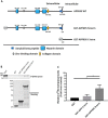
Matriptase interacts with APP770, APP751, and APP695. A, schematic representation of the GFP-tagged isoforms of APP. The structural elements of APP are depicted, including the heparin, KPI, OX-2, zinc-binding, collagen, amyloid β, and transmembrane domains. B, lysate of HEK293 cells transfected with matriptase and GFP-tagged APP770 (left panel), APP751 (middle panel), or APP695 (right panel) or with GFP were immunoprecipitated (IP) with GFP-Trap beads and then immunoblotted with anti-matriptase or anti-GFP antibodies to detect matriptase (Mat) and APP isoforms, respectively (n = 3 for each APP isoforms).
Figure 3.
Figure 3.
In vitro interaction of matriptase with the ectodomain of APP695. A, schematic representation of the GST-tagged APP695 deletion mutants used to determine the matriptase-binding domain. B, APP695 mutants and GST protein (10 μg) were immobilized on glutathione beads and incubated with in vitro translated 35S-labeled matriptase. Bound proteins were separated by SDS-PAGE and detected by autoradiography. GST proteins were detected with Coomassie Blue staining. Input, 2.5% of the total in vitro translated product (n = 6). C, a Kruskal–Wallis test on the densitometric analysis of B was applied. There is a statistical difference between GST alone and GST-APP695 N-term and between GST-APP695 C-term and GST-APP695 N-term (*, p < 0.05). Error bars represent means ± S.D.
Figure 4.
Figure 4.
Matriptase cleaves APP770, APP751, and APP695 in cellulo and in vitro. A, HEK293 cells were transfected with WT matriptase (Mat), a catalytically inactive matriptase mutant (S805A), or empty vector (Mock) together with GFP-tagged APP770 (left panel), APP751 (middle panel), or APP695 (right panel). Lysates and conditioned media of these cells were immunoblotted (IB) with anti-matriptase or anti-GFP antibody to detect matriptase, APP isoforms, and APP fragments (n = 3 for each isoform). Note the GFP-tagged APP fragment (cleaved) of 35 kDa in cell lysate and medium (arrow). B, HEK293 cells transfected with GFP-tagged APP770 (left panel), APP751 (middle panel), or APP695 (right panel) were incubated without (Buffer) or with 5 nm recombinant WT matriptase (sMat-WT) or catalytically inactive matriptase mutant (sMat-S805A). Conditioned media were immunoblotted as described in A. C, SH-SY5Y cells expressing endogenous APP were incubated without (Buffer) or with 5 nm of recombinant WT matriptase (sMat-WT) or catalytically inactive matriptase mutant (sMat-S805A). Conditioned media were immunoblotted with anti-APP N-terminal antibody (22C11) to detect APP and APP fragments in the medium (n = 3). Note the APP fragment (cleaved) of 10 kDa. D, in vitro translated 35S-labeled APP770 (left panel), APP751 (middle panel), and APP695 (right panel) were incubated with different concentrations (0, 1, 10, and 100 nm) of recombinant WT matriptase (sMat) or 100 nm catalytically inactive matriptase mutant (sS805A) for 1 h at 37 °C. Reaction products were separated by SDS-PAGE and detected by autoradiography (n = 3). Note the APP fragment (cleaved) of 10 kDa (arrow) and other higher molecular mass fragments (asterisks) in the presence of recombinant WT matriptase.
Figure 5.
Figure 5.
Matriptase cleaves APP at arginine 102. A, tandem mass spectrometry spectra from peptide ion trap collision-induced dissociation fragmentation for the peptide KQCKTHPHFVIPY identified after in vitro digestion and LC-MS/MS analysis of GST-APP695 ectodomain fragments generated by matriptase cleavage. Shown is a representative annotated MS/MS fragmentation spectrum with the identified matched N terminus-containing ions (b ions) in blue and the C terminus-containing ions (y ions) in red. Peptide intensities were summarized per amino acid residue and plotted in relation to each other. The detected peptide sequences indicate that Arg-102 is the main cleavage site. B, schematic representation of the position of the Arg cleaved by matriptase in the ectodomain of APP695 tagged with GST that was used to determine the matriptase cleavage site in A. C, HEK293 cells transfected with GFP-tagged APP695 wild type or in which Arg-102 was mutated to Ala (APP695 R102A) were incubated without (Buffer) or with 5 nm recombinant WT matriptase (sMat-WT). Conditioned media were immunoblotted (IB) with anti-GFP antibody to detect sAPP and APP fragments (n = 3). Note the absence of the GFP-tagged APP fragment (cleaved) of 35 kDa in the GFP-APP695 R102A lanes.
Figure 6.
Figure 6.
Matriptase cleavage of APP at Arg-102 alters Aβ40 production. A, SH-SY5Y cells were incubated without (Buffer) and with 5 nm recombinant WT matriptase (sMat-WT) or catalytically inactive matriptase mutant (sMat-S805A). Conditioned media were collected after 36 h, and Aβ40 levels were analyzed by ELISA. Error bars represent means ± S.D. Results are expressed as means ± S.D. (n = 3) and are normalized to the protein concentration in the medium. ***, p < 0.001. B, HEK293 cells transfected with GFP-tagged APP695 wild type or in which Arg-102 was mutated to Ala (APP695 R102A) and without or with matriptase (Mat). Aβ40 levels in the conditioned media were analyzed by ELISA as described in A. **, p < 0.05; ns, not significant.

References

    1. Chiang P. K., Lam M. A., and Luo Y. (2008) The many faces of amyloid β in Alzheimer's disease. Curr. Mol. Med. 8, 580–584 - PubMed
    1. Arai H., Lee V. M., Messinger M. L., Greenberg B. D., Lowery D. E., and Trojanowski J. Q. (1991) Expression patterns of β-amyloid precursor protein (β-APP) in neural and nonneural human tissues from Alzheimer's disease and control subjects. Ann. Neurol. 30, 686–693 - PubMed
    1. Kitaguchi N., Takahashi Y., Tokushima Y., Shiojiri S., and Ito H. (1988) Novel precursor of Alzheimer's disease amyloid protein shows protease inhibitory activity. Nature 331, 530–532 - PubMed
    1. Schmaier A. H., Dahl L. D., Rozemuller A. J., Roos R. A., Wagner S. L., Chung R., and Van Nostrand W. E. (1993) Protease nexin-2/amyloid β protein precursor. A tight-binding inhibitor of coagulation factor IXa. J. Clin. Investig. 92, 2540–2545 - PMC - PubMed
    1. Preece P., Virley D. J., Costandi M., Coombes R., Moss S. J., Mudge A. W., Jazin E., and Cairns N. J. (2004) Amyloid precursor protein mRNA levels in Alzheimer's disease brain. Brain Res. Mol. Brain Res. 122, 1–9 - PubMed

Publication types

MeSH terms

Associated data

LinkOut - more resources