Case Reports
. 2018 Jan;17(1):222-231.
doi: 10.1158/1535-7163.MCT-17-0472. Epub 2017 Oct 20.

ALK Inhibitor Response in Melanomas Expressing EML4-ALK Fusions and Alternate ALK Isoforms

Affiliations
Case Reports

ALK Inhibitor Response in Melanomas Expressing EML4-ALK Fusions and Alternate ALK Isoforms

Kasey L Couts et al. Mol Cancer Ther. 2018 Jan.

Abstract

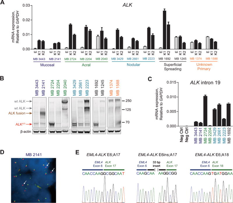
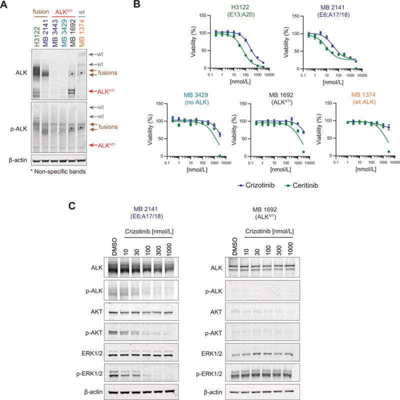
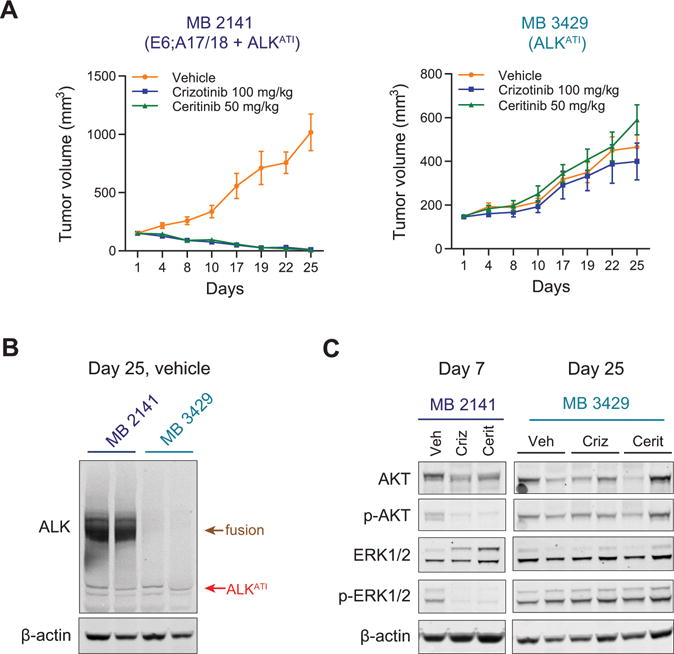
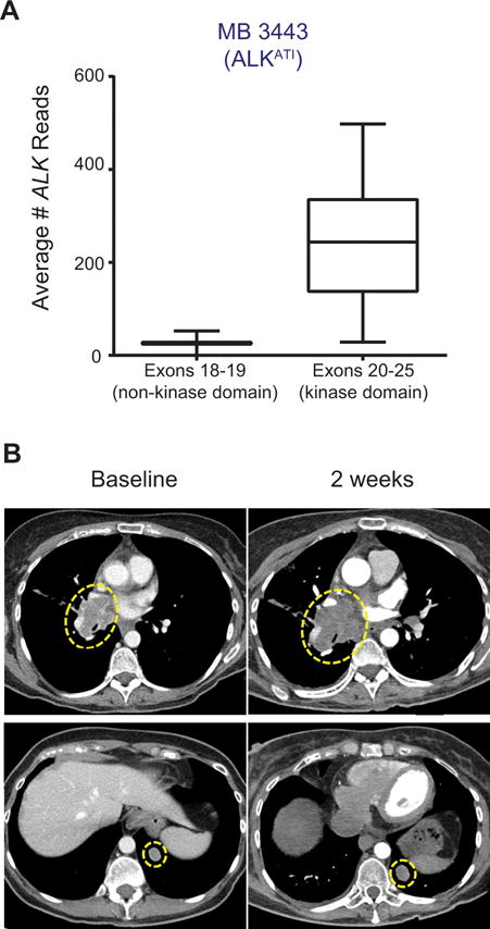
Oncogenic ALK fusions occur in several types of cancer and can be effectively treated with ALK inhibitors; however, ALK fusions and treatment response have not been characterized in malignant melanomas. Recently, a novel isoform of ALK (ALKATI ) was reported in 11% of melanomas but the response of melanomas expressing ALKATI to ALK inhibition has not been well characterized. We analyzed 45 melanoma patient-derived xenograft models for ALK mRNA and protein expression. ALK expression was identified in 11 of 45 (24.4%) melanomas. Ten melanomas express wild-type (wt) ALK and/or ALKATI and one mucosal melanoma expresses multiple novel EML4-ALK fusion variants. Melanoma cells expressing different ALK variants were tested for response to ALK inhibitors. Whereas the melanoma expressing EML4-ALK were sensitive to ALK inhibitors in vitro and in vivo, the melanomas expressing wt ALK or ALKATI were not sensitive to ALK inhibitors. In addition, a patient with mucosal melanoma expressing ALKATI was treated with an ALK/ROS1/TRK inhibitor (entrectinib) on a phase I trial but did not respond. Our results demonstrate ALK fusions occur in malignant melanomas and respond to targeted therapy, whereas melanomas expressing ALKATI do not respond to ALK inhibitors. Targeting ALK fusions is an effective therapeutic option for a subset of melanoma patients, but additional clinical studies are needed to determine the efficacy of targeted therapies in melanomas expressing wt ALK or ALKATIMol Cancer Ther; 17(1); 222-31. ©2017 AACR.


Figures

Figure 1
Figure 1
Identification of melanoma tumors expressing wt ALK, ALKATI, and an EML4-ALK fusion. A, ALK mRNA expression was quantified in melanoma PDX tumors by qRT-PCR analysis with primers located in early exons (E) or exons within the kinase domain (K1, K2) of ALK. Expression was normalized to GAPDH and error bars represent SEM of triplicate reactions. The 12 ALK expression positive tumors out of 45 total PDX tumors analyzed are shown. B, Immunoblot analysis of ALK positive PDX tumor lysates. C, Expression of ALK intron 19 was quantified by qRT-PCR. Expression was normalized to GAPDH and error bars represent SEM of triplicate reactions. D, Fluorescence in-situ hybridization (FISH) analysis of the MB 2141 PDX tumor using ALK specific 5′ (green) and 3′ (red) break-apart probes. E, Sanger sequencing analysis of EML4-ALK fusion variant transcripts. PCR products were generated from MB 2141 PDX cDNA using an EML4-exon 4 forward primer and ALK exon 17 or 20 reverse primers. PCR products were sub-cloned and analyzed by Sanger sequencing.
Figure 2
Figure 2
ALK inhibitors reduce viability in cells expressing EML4-ALK fusions, but not in cells expressing ALKATI or wt ALK. A, Immunoblot analysis of lysates H3122 NSCLC cells and melanoma cells. B, Cells were treated with increasing concentrations of ALK inhibitors crizotinib or ceritinib. Viability was analyzed after 72 hours and normalized to DMSO treated controls. C, Cells were treated with increasing concentrations of crizotinib for 2 hours and analyzed by immunoblotting.
Figure 3
Figure 3
Melanoma cells respond to targeted MEK inhibition against other oncogenic drivers. A, MB 1692, MB 3443, and MB 3429 cells were treated with increasing concentrations of a MEK inhibitor (trametinib) or mutant BRAF specific inhibitor (vemurafenib). Viability was analyzed after 72 hours and normalized to DMSO treated controls. B, Cells were treated with increasing concentrations of trametinib for 2 hours and analyzed by immunoblotting. C, MB 1692 cells were treated with increasing concentrations of ceritinib alone or in combination with 1 nmol/L or 2 nmol/L trametinib. Viability was analyzed after 72 hours and normalized to non-treated controls. D, MB 1692 cells were treated with the indicated concentrations of trametinib and/or ceritinib for 2 hours and analyzed by immunoblotting. For all viability results, error bars represent SEM of three technical triplicates.
Figure 4
Figure 4
ALK inhibitors decrease growth of melanoma xenograft tumors expressing an EML4-ALK fusion, but not ALKATI only. A, MB 2141 and MB 1692 subcutaneous xenograft tumors were generated in nude mice and treated with vehicle, crizotinib (100 mg/kg), or ceritinib (50 mg/kg). Growth was measured bi-weekly for 25 days, at which time all tumors were harvested. B, Immunoblot analysis for total ALK and β-actin in tumor lysates from vehicle tumors harvested at day 25. Lysates from two independent tumors were analyzed for each condition. C, Immunoblot analysis for AKT/p-AKT, ERK1/2/p-ERK/1/2, and β-actin in MB 2141 tumors harvested after 7 days of treatment and MB 3429 tumors harvested after 25 days of treatment. Lysates from two independent tumors were analyzed for each condition for MB 3429 tumors. (Veh, vehicle; Criz, crizotinib; Cerit, ceritinib).
Figure 5
Figure 5
A patient with melanoma expressing ALKATI does not respond to treatment with entrectinib. A, The patient specimen was analyzed for ALK gene expression using targeted RNA next-generation sequencing. The average number of sequencing reads for exons 18–19 (upstream of the kinase domain, not included in ALKATI) and exons 20–25 (kinase domain included in ALKATI) of the ALK gene are shown. B, Radiographic images from CT scans performed prior to treatment (baseline) and 2 weeks after the start of treatment are shown for a right hilar pulmonary tumor nodule (top panels) and a left lower lobe pulmonary tumor nodule (bottom panels).

Similar articles

Cited by

References

    1. Soda M, Choi YL, Enomoto M, Takada S, Yamashita Y, Ishikawa S, et al. Identification of the transforming EML4-ALK fusion gene in non-small-cell lung cancer. Nature. 2007;448:561–6. - PubMed
    1. Aisner DL, Nguyen TT, Paskulin DD, Le AT, Haney J, Schulte N, et al. ROS1 and ALK fusions in colorectal cancer, with evidence of intratumoral heterogeneity for molecular drivers. Mol Cancer Res. 2014;12:111–8. - PMC - PubMed
    1. Lin E, Li L, Guan Y, Soriano R, Rivers CS, Mohan S, et al. Exon array profiling detects EML4-ALK fusion in breast, colorectal, and non-small cell lung cancers. Mol Cancer Res. 2009;7:1466–76. - PubMed
    1. Lawrence B, Perez-Atayde A, Hibbard MK, Rubin BP, Dal Cin P, Pinkus JL, et al. TPM3-ALK and TPM4-ALK oncogenes in inflammatory myofibroblastic tumors. Am J Pathol. 2000;157:377–84. - PMC - PubMed
    1. Kwak EL, Bang YJ, Camidge DR, Shaw AT, Solomon B, Maki RG, et al. Anaplastic lymphoma kinase inhibition in non-small-cell lung cancer. N Engl J Med. 2010;363:1693–703. - PMC - PubMed

Publication types

Substances