. 2018 Jan;17(1):254-263.
doi: 10.1158/1535-7163.MCT-16-0868. Epub 2017 Oct 20.

AKT1low Quiescent Cancer Cells Promote Solid Tumor Growth

Affiliations

AKT1low Quiescent Cancer Cells Promote Solid Tumor Growth

Cleidson P Alves et al. Mol Cancer Ther. 2018 Jan.

Abstract

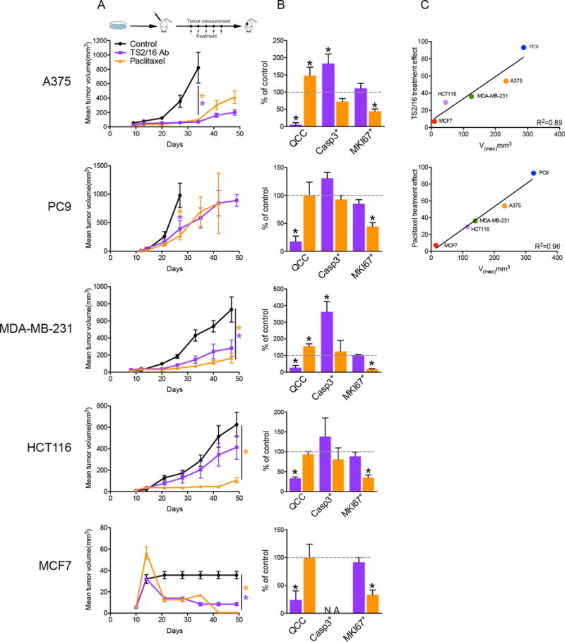
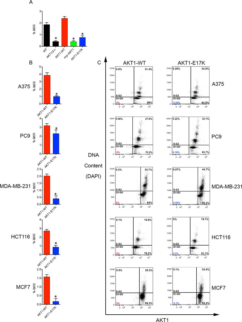
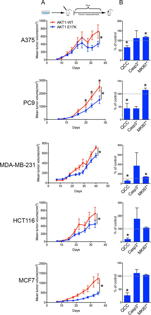
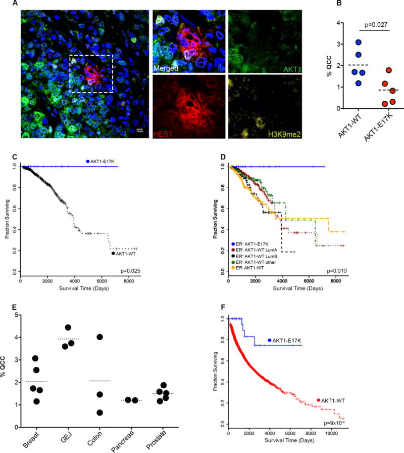
Human tumor growth depends on rapidly dividing cancer cells driving population expansion. Even advanced tumors, however, contain slowly proliferating cancer cells for reasons that remain unclear. Here, we selectively disrupt the ability of rapidly proliferating cancer cells to spawn AKT1low daughter cells that are rare, slowly proliferating, tumor-initiating, and chemotherapy-resistant, using β1-integrin activation and the AKT1-E17K-mutant oncoprotein as experimental tools in vivo Surprisingly, we find that selective depletion of AKT1low slow proliferators actually reduces the growth of a molecularly diverse panel of human cancer cell xenograft models without globally altering cell proliferation or survival in vivo Moreover, we find that unusual cancer patients with AKT1-E17K-mutant solid tumors also fail to produce AKT1low quiescent cancer cells and that this correlates with significantly prolonged survival after adjuvant treatment compared with other patients. These findings support a model whereby human solid tumor growth depends on not only rapidly proliferating cancer cells but also on the continuous production of AKT1low slow proliferators. Mol Cancer Ther; 17(1); 254-63. ©2017 AACR.


Figures

Fig. 1
Fig. 1. Activation of β1-integrin reduces QCCs and tumor growth in vivo
Mice with subcutaneous tumors were treated with either TS2/16 monoclonal antibody (18 mg/kg, i.p.) (purple) or paclitaxel (20 mg/kg, i.p.) (gold) once a week for 5 weeks versus untreated control (black) in 5 different xenograft models (n = 8 mice/arm). Tumor growth (A) and bar graph percentages of QCCs, cleaved-Caspase3+, and MKI67+ cells in TS2/16-treated (purple) or paclitaxel-treated (gold) versus untreated control tumors (B) in different xenograft models is shown. Error bars indicate mean ± SEM, * = p < 0.05. (C) Correlation of maximum velocity of tumor growth (Vmax) versus treatment effect for TS2/16 and paclitaxel.
Fig. 2
Fig. 2. AKT1-E17K disrupts QCCs
(A) Bar graph percentages of QCCs in the HCT116-AKT1/2−/− human cancer cell line with cDNAs for AKT1-WT or AKT1 mutants. (B) Bar graph percentages of QCCs in human cancer cell lines ectopically expressing inducible AKT1-WT or AKT1-E17K. Error bars indicate mean ± SEM for three independent experiments, * = p < 0.05. (C) Flow cytometry analysis of cell cycle (DAPI) and AKT1 levels.
Fig. 3
Fig. 3. AKT1-E17K reduces QCCs and tumor growth in vivo
Human cancer cells ectopically expressing inducible AKT1-WT or AKT1-E17K were injected into mice, with induction two days after injection, in 5 different xenograft models (n = 10 mice/arm). Tumor growth (A) and bar graph percentages of QCCs, cleaved-Caspase3+, and MKI67+ cells in AKT1-E17K versus AKT1-WT tumors (B) in different xenograft models is shown. Error bars indicate mean ± SEM, * = p < 0.05.
Fig. 4
Fig. 4. AKT1-E17K, QCCs, and clinical outcome in human cancers
(A) Illustrative image of QCC in single section from a human ER+ primary breast tumor (Blue = DAPI nuclear stain). (B) percentage of QCCs in individual human breast tumors. (C) Kaplan–Meier plots of overall survival in ER+/AKT1-E17K breast cancer patients versus patients with ER+/AKT1- WT breast tumors. (D) ER+/AKT1-WT breast tumors subdivided by known molecular subtypes (from TCGA). (E) Percentage of QCCs in individual primary tumors from a variety of epithelial cancer types (i.e., breast, gastro-esophageal junction, colon, pancreas, prostate). (F) Kaplan–Meier plot of overall survival in all cancer patients with AKT1-E17K mutation versus AKT1-WT (from TCGA).

References

    1. Loddo M, Kingsbury SR, Rashid M, Proctor I, Holt C, Young J, et al. Cell-cycle-phase progression analysis identifies unique phenotypes of major prognostic and predictive significance in breast cancer. British journal of cancer. 2009;100:959–70. - PMC - PubMed
    1. Yeh AC, Ramaswamy S. Mechanisms of cancer cell dormancy - Another hallmark of cancer? Cancer research. 2015;75:5014–22. - PMC - PubMed
    1. Nowell PC. The clonal evolution of tumor cell populations. Science. 1976;194:23–8. - PubMed
    1. Massague J. G1 cell-cycle control and cancer. Nature. 2004;432:298–306. - PubMed
    1. Dey-Guha I, Wolfer A, Yeh AC, J GA, Darp R, Leon E, et al. Asymmetric cancer cell division regulated by AKT. Proceedings of the National Academy of Sciences of the United States of America. 2011;108:12845–50. - PMC - PubMed

Publication types

Substances