. 2018 Mar;14(3):306-317.
doi: 10.1016/j.jalz.2017.09.008. Epub 2017 Oct 19.

Amyloid β synaptotoxicity is Wnt-PCP dependent and blocked by fasudil

Affiliations

Amyloid β synaptotoxicity is Wnt-PCP dependent and blocked by fasudil

Katherine J Sellers et al. Alzheimers Dement. 2018 Mar.

Abstract

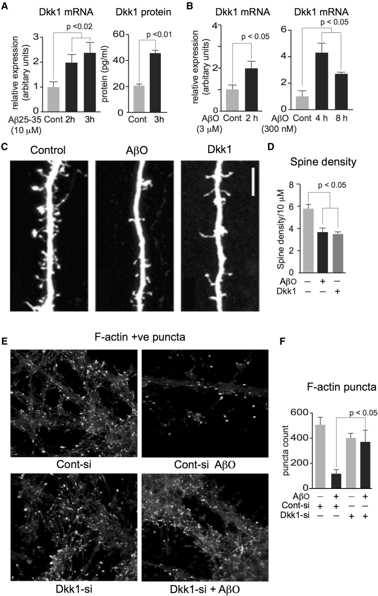
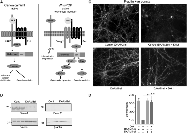
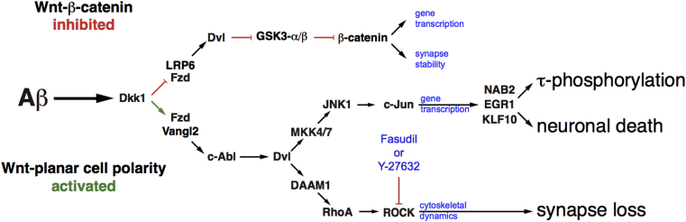
Introduction: Synapse loss is the structural correlate of the cognitive decline indicative of dementia. In the brains of Alzheimer's disease sufferers, amyloid β (Aβ) peptides aggregate to form senile plaques but as soluble peptides are toxic to synapses. We previously demonstrated that Aβ induces Dickkopf-1 (Dkk1), which in turn activates the Wnt-planar cell polarity (Wnt-PCP) pathway to drive tau pathology and neuronal death.

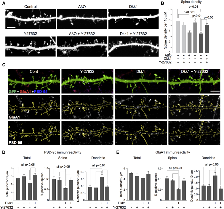
Methods: We compared the effects of Aβ and of Dkk1 on synapse morphology and memory impairment while inhibiting or silencing key elements of the Wnt-PCP pathway.

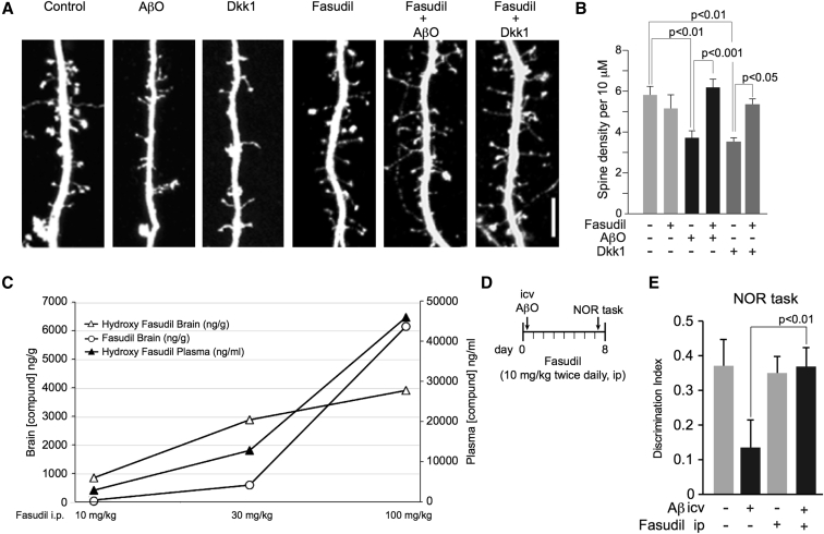
Results: We demonstrate that Aβ synaptotoxicity is also Dkk1 and Wnt-PCP dependent, mediated by the arm of Wnt-PCP regulating actin cytoskeletal dynamics via Daam1, RhoA and ROCK, and can be blocked by the drug fasudil.

Discussion: Our data add to the importance of aberrant Wnt signaling in Alzheimer's disease neuropathology and indicate that fasudil could be repurposed as a treatment for the disease.

Keywords: Alzheimer's disease; Amyloid; DAAM1; Dickkopf-1; Fasudil; Planar cell polarity; ROCK; Synapse; Synaptotoxicity; Wnt.


Figures

Fig. 1
Fig. 1
Aβ synaptotoxicity is Dkk1 dependent. (A) Rat primary cortical neuronal cultures (14 d.i.v.) were treated with 10 μM Aβ25–35 for 2 and 3 hours, cells were harvested for RNA extraction, and media were collected for protein analysis. cDNA was generated and qRT-PCR performed to determine rat Dkk1 mRNA levels, left. Secreted Dkk1 protein levels in media were measured by ELISA, right. (B) Similar cultures were treated at 3 μM and 300 nM with AβO preparation for the times indicated and harvested, and Dkk1 mRNA levels were determined as mentioned previously. (C and D) Similar cultures were transfected with eGFP at 24 d.i.v., 48 hours later treated for 4 hours with 2 μM AβO, or for 3 hours with 400 ng/mL Dkk1, fixed, imaged by confocal microscopy, and dendritic spine density and morphology assessed. Both treatments resulted in a significant reduction in dendritic spine linear density, quantified in (D), scale bar = 5 μM. Dendritic spine linear density/10 μm: control, 5.8 ± 0.41; AβO, 3.7 ± 0.34; Dkk1, 3.5 ± 0.31; P < .001 for all treatments. (E and F) Similar cultures were treated overnight with Dkk1-siRNA duplex, or a scrambled version as control, each linked to the Pen-1 peptide. Next day, cells were treated with 2 μM AβO for 4 hours, fixed, and fluorescently labeled with phalloidin-488, imaged (E), and F-actin–labeled puncta quantified (F), scale bar = 50 μM. In all the aforementioned, significance was determined by ANOVA and Tukey's post hoc t-test. Error bars indicate standard error of the mean. Abbreviations: AβO, amyloid β (1–42) oligomers; ANOVA, analysis of variance; d.i.v., days in vitro; eGFP, enhanced green fluorescent protein; Dkk1, Dickkopf-1.
Fig. 2
Fig. 2
Dkk1 synaptotoxicity is Daam1 dependent. (A) Schematic of the Wnt-PCP pathway, showing the two arms branching below disheveled, acting via Daam/Rho/ROCK to regulate cytoskeletal dynamics and JNK/c-Jun primarily to regulate gene transcription. (B) Primary cortical neuronal cultures were treated with DAAM1 or DAAM2 Pen-1–coupled siRNA duplexes for 48 hours, harvested, and analyzed by Western blotting for Daam1 and Daam2. Daam1 was detectable in untreated cells, whereas Daam2 was not. Daam1-si potently reduced Daam1 protein expression levels. (C and D) Cultures were treated overnight with DAAM1- or DAAM2-siRNA duplexes. Next day, cells were treated with 400 ng/mL recombinant Dkk1 protein for 3 hours, fixed, and fluorescently labeled with phalloidin-488, imaged (C), scale bar = 50 μM, and F-actin–labeled puncta quantified (D). Significance determined by ANOVA and Tukey's post hoc t-test. Error bars indicate standard deviation. Abbreviations: Aβ, amyloid β; ANOVA, analysis of variance; c-Abl1, c-Abl oncogene 1, nonreceptor tyrosine kinase (ABL1); d.i.v., days in vitro; DAAM1, disheveled associated activator of morphogenesis 1; Dkk1, Dickkopf-1; Dvl, disheveled; EGR1, early growth response 1; Fzd, frizzled; GSK3-α/β, glycogen synthase kinase-α/β; JNK1, c-Jun N-terminal kinase (MAPK8); KLF10, Krüppel-like factor 10; LRP6, low-density lipoprotein receptor–related protein 6; MKK4/7, mitogen-activated protein kinase 4/7 (MAP2K4 and MAP2K7); NAB2, NGFI-A binding protein 2; PCP, planar cell polarity; RhoA, Ras homolog family member A; ROCK, Rho-associated coiled-coil containing protein kinase; Vangl2, Van Gogh–like protein 2.
Fig. 3
Fig. 3
Aβ and Dkk1 synaptic effects are ROCK dependent. (A and B) Rat primary cortical neurons were transfected with eGFP at 24 d.i.v. and 48 hours later treated with Y-27632 or vehicle, and 15 minutes later with 2 μM AβO for 4 hours or 400 ng/mL Dkk1 for 3 hours, fixed, and imaged by confocal microscopy for the examination of spine morphology. AβO and Dkk1 caused a significant reduction in dendritic spine linear density. Y2763 alone had no significant effect on spine density, but in combination with AβO and Dkk1, it blocked the effect of both (scale bar = 5 μM). (C, D, and E) Rat primary 26-d.i.v. neurons expressing eGFP were treated with Y-27632 and Dkk1, all as mentioned previously (3A). (D) Concurrent with a loss of spine density, Dkk1 caused a significant reduction in total PSD-95 puncta (Total PSD-95 density/10 μm: control, 5.5 ± 0.34; control + Y-27632, 6.2 ± 0.55; Dkk1, 4.3 ± 0.39; Dkk1 + Y-27632, 6.6 ± 0.43). Interestingly, the number of spines containing PSD-95 was also reduced, with a concurrent increase in the density of dendritic PSD-95, following treatment with Dkk1. This effect was blocked by Y-27632 (% spines containing PSD-95: control, 79.8 ± 2.4; control + Y-27632, 78.2 ± 2.7; Dkk1, 59.1 ± 3.7; Dkk1 + Y-27632, 78.9 ± 2.1). Dendritic PSD-95 puncta/10 μm: control, 0.93 ± 0.11; control + Y-27632, 0.89 ± 0.11; Dkk1, 1.67 ± 0.17; Dkk1 + Y-27632, 0.95 ± 0.15. (E) Dkk1 did not significantly affect the total level of GluA1-immunoreactive puncta but did reduce the number of spines positive for GluA1 and increased levels of GluA1 in dendrites, which was again blocked by inhibition of ROCK. (1) GluA1 linear density/10 μm: control, 5.0 ± 0.34; control + Y-27632, 4.6 ± 0.40; Dkk1, 4.2 ± 0.40; Dkk1 + Y-27632, 4.9 ± 0.44; P = .4574; (2) % spines containing GluA1: control, 67.8 ± 4.3; control + Y-27632, 68.4 ± 3.7; Dkk1, 47.9 ± 4.4; Dkk1 + Y-27632, 65.6 ± 3.1; and (3) Dendritic GluA1 puncta/10 μm: control, 1.25 ± 0.13; control + Y-27632, 1.20 ± 0.15; Dkk1, 2.09 ± 0.26; Dkk1 + Y-27632, 0.99 ± 0.22. In all bar graphs, error bars indicate standard error of the mean. Abbreviations: AβO, amyloid β (1–42) oligomers; Dkk1, Dickkopf-1; eGFP, enhanced green fluorescent protein; ROCK, Rho-associated coiled-coil containing protein kinase.
Fig. 4
Fig. 4
Fasudil is CNS penetrant and blocks Aβ synaptotoxicity and cognitive impairment. (A and B) Rat primary cortical neuronal cultures were transfected with eGFP at 26 d.i.v. and 48 hours later pretreated with 5 μM fasudil or vehicle for 15 minutes and subsequently treated with AβO or Dkk1 and imaged as mentioned in Fig. 3A, scale bar = 5 μM. AβO and Dkk1 caused a significant reduction in dendritic spine linear density, which fasudil fully and significantly blocked, as shown in (B) (dendritic spine linear density/10 μm: control, 5.8 ± 0.41; fasudil, 5.2 ± 0.64; AβO, 3.7 ± 0.34; AβO + fasudil, 6.2 ± 0.38; Dkk1, 3.5 ± 0.20; Dkk1 + fasudil, 5.4 ± 0.27). (B) Significance determined by ANOVA and Tukey's post hoc t-test. In (B), error bars indicate standard error of the mean. (C) Male CD1 mice were administered fasudil or hydroxyfasudil at 10, 30, and 100 mg/kg, ip, and brain and plasma were collected 20 minutes after injection. Fasudil and hydroxyfasudil were detected and measured by mass spectrometry. Plasma levels of fasudil were below the threshold of detection at all doses, and its data points were omitted from the graph. (D and E) 40 young adult female rats were administered 10 mg/kg fasudil, or vehicle, ip, twice daily for 7 days, and given either a single dose of AβO or vehicle, unilaterally, icv, on day 1. On day 7, all animals were presented with an NOR task, schematized in (D) Rats receiving vehicle and AβO showed profound deficit in this task, while the performance of rats receiving AβO and fasudil was not different to that of controls (E) Error bars in (E) indicate standard deviation. Abbreviations: AβO, amyloid β (1–42) oligomers; Dkk1, Dickkopf-1; NOR, novel object recognition; eGFP, enhanced green fluorescent protein; icv, intracerebroventricularly; ip, intraperitoneally.
Fig. 5
Fig. 5
Schematic of Aβ-driven Wnt-PCP pathway activation. Aβ drives a rapid increase in Dkk1 expression. Concomitant with antagonism of canonical Wnt-β-catenin–signaling Dkk1 then drives the activation of the Wnt-PCP pathway by antagonizing the LRP6-Fzd interaction. We have previously shown that activity in the JNK/c-Jun arm of Wnt-PCP induces the expression of several identified genes required for Aβ-driven increases in tau phosphorylation and neuronal death to occur. Here, we demonstrate that activity of the Daam1/RhoA/ROCK arm is necessary for Aβ-driven synaptotoxicity and that this can be blocked by ROCK inhibitors Y-27632 or fasudil. Abbreviations: Aβ, amyloid β; c-Abl1, c-Abl oncogene 1, nonreceptor tyrosine kinase (ABL1); DAAM1, disheveled associated activator of morphogenesis 1; Dkk1, Dickkopf-1; Dvl, disheveled; EGR1, early growth response 1; Fzd, frizzled; GSK3-α/β, glycogen synthase kinase-α/β; JNK1, c-Jun N-terminal kinase (MAPK8); KLF10, Krüppel-like factor 10; LRP6, low-density lipoprotein receptor–related protein 6; MKK4/7, mitogen-activated protein kinase 4/7 (MAP2K4 and MAP2K7); NAB2, NGFI-A binding protein 2; PCP, planar cell polarity; RhoA, Ras homolog family member A; ROCK, Rho-associated coiled-coil containing protein kinase; Vangl2, Van Gogh–like protein 2.

References

    1. Vargas L.M., Leal N., Estrada L.D., Gonzalez A., Serrano F., Araya K. EphA4 activation of c-Abl mediates synaptic loss and LTP blockade caused by amyloid-beta oligomers. PLoS One. 2014;9:e92309. - PMC - PubMed
    1. Pozueta J., Lefort R., Shelanski M.L. Synaptic changes in Alzheimer's disease and its models. Neuroscience. 2013;251:51–65. - PubMed
    1. Lambert M.P., Barlow A.K., Chromy B.A., Edwards C., Freed R., Liosatos M. Diffusible, nonfibrillar ligands derived from Aβ1–42 are potent central nervous system neurotoxins. Proc Natl Acad Sci U S A. 1998;95:6448–6453. - PMC - PubMed
    1. Killick R., Ribe E.M., Al-Shawi R., Malik B., Hooper C., Fernandes C. Clusterin regulates beta-amyloid toxicity via Dickkopf-1-driven induction of the wnt-PCP-JNK pathway. Mol Psychiatry. 2014;19:88–98. - PMC - PubMed
    1. Bafico A., Liu G., Yaniv A., Gazit A., Aaronson S.A. Novel mechanism of Wnt signalling inhibition mediated by Dickkopf-1 interaction with LRP6/Arrow. Nat Cell Biol. 2001;3:683–686. - PubMed

Publication types

MeSH terms