Review
. 2018 Mar;26(3):191-208.
doi: 10.1016/j.tim.2017.09.012. Epub 2017 Oct 19.

The Molecular Basis of Noncanonical Bacterial Morphology

Affiliations
Review

The Molecular Basis of Noncanonical Bacterial Morphology

Paul D Caccamo et al. Trends Microbiol. 2018 Mar.

Abstract

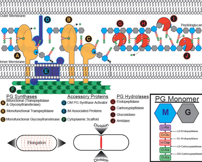
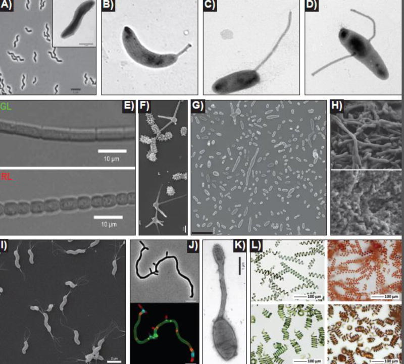
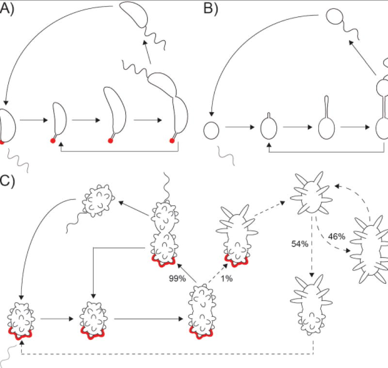
Bacteria come in a wide variety of shapes and sizes. The true picture of bacterial morphological diversity is likely skewed due to an experimental focus on pathogens and industrially relevant organisms. Indeed, most of the work elucidating the genes and molecular processes involved in maintaining bacterial morphology has been limited to rod- or coccal-shaped model systems. The mechanisms of shape evolution, the molecular processes underlying diverse shapes and growth modes, and how individual cells can dynamically modulate their shape are just beginning to be revealed. Here we discuss recent work aimed at advancing our knowledge of shape diversity and uncovering the molecular basis for shape generation in noncanonical and morphologically complex bacteria.

Keywords: bacterial shape; morphological engineering; morphology; peptidoglycan; pleomorphism.


Figures

Figure 1
Figure 1. A simplified accounting of peptidoglycan remodeling components in Gram-negative bacteria
Due to the scope of this review we will only briefly describe common themes in the proteins involved in PG remodeling, using Gram-negatives as an example (for detailed reviews regarding PG remodeling enzymes, start with [7, 11, 148]). Cartoons are not meant to imply an experimentally determined structure for the proteins. Adapted in part from [11]. Inset, right: structure of uncrosslinked PG monomer depicting the disaccharide N-Acetylmuramic acid ("M") and N-Acetylglucosamine ("G") and the pentapeptide stem, from proximal to distal, L-alanine ("L-Ala"), D-glutamic acid ("D-Glu"), meso-diaminopimelic acid ("m-Dap"; a derivative of lysine), and two D-alanines ("D-Ala"). From proximal to distal, the stem peptide isoform pattern is "L-D-L-D-D". Enzymes that break bonds between the stem peptides are prefixed by the isoforms for the peptides they separate. The "makers", or (A–C) PG synthases, assemble the nascent PG meshwork. (A&C) Glycosyltransferases polymerize PG monomers into glycan strands, while (A&B) transpeptidases form crosslinks between the stem peptides to form the sacculus. (D–F) Accessory and SEDS proteins include: (D) Outer membrane anchored PG synthase activators and (E) inner membrane (IM) associated proteins. (E) IM associated proteins include enzymes that synthesize PG monomers in the cytoplasm and flippases that flip the monomers across the IM to the periplasm, SEDS family proteins [–16], and proteins that help anchor components on the PG synthesis machinery to (F) cytoplasmic scaffolding proteins. (F) Scaffolding proteins recruit various cytoplasmic and IM proteins associated with PG synthesis and localize synthesis activity. The "breakers", or (G–J) PG hydrolases, modify PG after synthesis. (G) Endopeptidases can break crosslinks ("DD-endopeptidases") or peptide linkages ("LD-endopeptidases" or "DL-endopeptidases") of non-terminal amino acids. (H) Carboxypeptidases trim the terminal stem peptide. Shown is removal of the terminal, fifth position D-Ala. (I) Glucosidases, of which there are different classes depending on which bond is broken and the type of catalytic reaction used, cleave the glycan strands. (J) Amidases remove the peptide stem from N-Acetylmuramic acid ("M", inset) in the glycan chain. Bottom, cell growth and division typical of E. coli: Dispersed growth along the long axis elongates the cell (left, dashed red lines), and a division septum (right, solid red ring) is formed at the midcell allowing the daughter cells to recapitulate the initial shape and size of the mother cell.
Figure 2
Figure 2. Diverse bacterial morphologies
A) Uncharacterized spiral-shaped methanotroph. Phase contrast with inset electron micrograph. Adapted from [24]. B)Caulobacter crescentus. Single polar prostheca [32]. C)Asticcacaulis excentricus. Single sub-polar prostheca [32]. D)Asticcacaulis biprosthecum. Two bilateral prosthecae [32]. E)Fremyella diplosiphon. Complimentary chromatic adaptation (CCA) mediated pleomorphism. Cells grown in green light (GL, top) are elongated and rectangular. Cells grown in red light (RL, bottom) are short and rounded. Adapted from [149]. F)Prosthecomicrobium hirschii. Electron micrograph showing short- and long-prosthecate morphotypes. Adapted from [73]. G)Dinoroseobacter shibae. Scanning electron micrograph showing the inherent morphological heterogeneity of wild-type D. shibae. Scale bar = 5 µm. [77]. H)Lactococcus lactis. Scanning electron micrograph showing different regions of the same L. lactis biofilm. The upper region of the biofilm contains elongated rods (top), while the lower region contains ovoid cells (bottom). Adapted from [60]. I)Helicobacter pylori. Scanning electron microscope images of wild-type H. pylori. Adapted from [96]. J)Streptomyces venezuelae. Virtual time-lapse of polarly growing S. venezuelae labeled with a long pulse (cell body) of green fluorescent D-amino acid (FDAA), followed by sequential short pulses of orange, blue, and red FDAAs (apical tips). Top = phase, bottom = fluorescence, scale bar = 5 µm. (Image courtesy of Yen-Pang Hsu, Indiana University). K)Hyphomonas adhaerens (related to H. neptunium). The mother cell (bottom), the prostheca (middle), and the developing daughter bud (top) are visible. Adapted from [150]. L)Spirulina (Arthrospira platensis). Spirulina biotemplated microcoils. The helical pitch of the cyanobacteria can be modified by tuning the culture conditions (left panels). Copper microcoils are produced through a electroless plating technique using Spirulina as the template (right panels). Adapted from [121].
Figure 3
Figure 3. Multimorphic life cycles of prosthecate Alphaproteobacteria
A) Dimorphic life cycle of Caulobacter crescentus. The prosthecate mother cell produces an adhesive holdfast (shown in red) at the tip of the prostheca. Cell division results in a motile, non-replicating swarmer cell that differentiates into a prosthecate cell. B) Dimorphic life cycle of Hyphomonas neptunium. The prosthecate mother cell produces a bud at the distal end of the prostheca. Upon septation, a motile, non-replicating swarmer cell is released that differentiates into a prosthecate cell. C) Multimorphic life cycle of Prosthecomicrobium hirschii. Most of the time, short-prosthecate P. hirschii cells follow a C. crescentus-like life cycle (solid arrows): a unipolar polysaccharide (UPP, an adhesin similar to C. crescentus holdfast; shown in red) producing mother cell gives rise to a motile, non-replicating swarmer cell that differentiates into a short-prosthecate cell. A rare event, short-prosthecate mother cells can give rise to non-motile, long-prosthecate cells that do not produce UPP (dashed arrow). Long-prosthecate mother cells can produce either long-prosthecate cells or short-prosthecate cells at roughly equal frequency (dashed arrows), though it should be noted that the observed frequency of morphotype conversion was observed on MMB (minimal medium broth) agarose pads and might differ in other conditions.

References

    1. Young KD. The selective value of bacterial shape. Microbiol Mol Biol Rev. 2006;70:660–703. - PMC - PubMed
    1. Kysela DT, et al. Diversity Takes Shape: Understanding the Mechanistic and Adaptive Basis of Bacterial Morphology. PLoS Biol. 2016;14:e1002565. - PMC - PubMed
    1. Yang DC, et al. Staying in Shape: the Impact of Cell Shape on Bacterial Survival in Diverse Environments. Microbiol Mol Biol Rev. 2016;80:187–203. - PMC - PubMed
    1. van Teeseling MCF, et al. Determinants of Bacterial Morphology: From Fundamentals to Possibilities for Antimicrobial Targeting. Front Microbiol. 2017;8:1264. - PMC - PubMed
    1. Allan EJ, et al. Bacterial L-forms. Adv Appl Microbiol. 2009;68:1–39. - PubMed

Publication types

LinkOut - more resources