. 2017 Nov 2;171(4):771-782.e11.
doi: 10.1016/j.cell.2017.09.051. Epub 2017 Oct 19.

The Human Knockout Gene CLYBL Connects Itaconate to Vitamin B12

Affiliations

The Human Knockout Gene CLYBL Connects Itaconate to Vitamin B12

Hongying Shen et al. Cell. .

Abstract

CLYBL encodes a ubiquitously expressed mitochondrial enzyme, conserved across all vertebrates, whose cellular activity and pathway assignment are unknown. Its homozygous loss is tolerated in seemingly healthy individuals, with reduced circulating B12 levels being the only and consistent phenotype reported to date. Here, by combining enzymology, structural biology, and activity-based metabolomics, we report that CLYBL operates as a citramalyl-CoA lyase in mammalian cells. Cells lacking CLYBL accumulate citramalyl-CoA, an intermediate in the C5-dicarboxylate metabolic pathway that includes itaconate, a recently identified human anti-microbial metabolite and immunomodulator. We report that CLYBL loss leads to a cell-autonomous defect in the mitochondrial B12 metabolism and that itaconyl-CoA is a cofactor-inactivating, substrate-analog inhibitor of the mitochondrial B12-dependent methylmalonyl-CoA mutase (MUT). Our work de-orphans the function of human CLYBL and reveals that a consequence of exposure to the immunomodulatory metabolite itaconate is B12 inactivation.

Keywords: C5 metabolism; CLYBL; citramalyl-CoA lyase; human genetics; itaconate; metabolism; mitochondria; systems biology; vitamin B(12).


Figures

Figure 1
Figure 1. Enzymatic activity and crystal structure of CLYBL
(A) Putative enzymatic activities of CLYBL. We detected (1) malate/methylmalate/citramalate synthase activity, (2) citramalyl-CoA lyase activity and (3) malyl-CoA thioesterase activity. (B) In vitro enzyme analysis of CLYBL reveals high citramalyl-CoA lyase activity. (C) Crystal structure of human CLYBL trimer. One subunit is shown in pink, magnesium ion in magenta, and the substrate mimic propionyl-CoA in cyan. The critical residue for catalytic activity Asp320 is highlighted. (D) Docking of citramalyl-CoA in the substrate binding pocket of the CLYBL structure. Free CoASH in the structure is shown in green, docked citramalyl-CoA in blue, one CLYBL subunit in pink and the neighboring subunit in gray. (E) The distances between docked citramalyl-CoA and the CLYBL residues are too close to accommodate additional carbon atoms in substrates larger than citramalyl-CoA. Three different views are shown. Labeled distances are in Angstrom (Å). The substrate-binding pocket of CLYBL is shown in surface representation.
Figure 2
Figure 2. CLYBL is a citramalyl-CoA lyase operating in C5 dicarboxylate catabolism
(A) Citramalyl-CoA levels in extracts from control and Clybl KO brown adipocytes. All results are means ± SD (n ≥ 3 biologic replicates) and were confirmed in independent experiments. (B) Citramalyl-CoA levels in the control brown adipocytes treated with 2 mM itaconate. (C) Schematic of the proposed CLYBL-mediated reaction in itaconate metabolism. (D) Schematic of itaconate -13C5 labeling into citramalyl-CoA. (E) – (F) Relative abundances of different mass isotopomers of cellular itaconate (in E) and citramalyl-CoA (in F) from control brown adipocytes treated with either labeled and unlabeled itaconate.
Figure 3
Figure 3. Loss of CLYBL leads to a cell-autonomous defect in mitochondrial B12 metabolism
(A) Western blot of Clybl in Clybl CRISPR KO murine brown adipocytes and Clybl KO cells expressing FLAG-tagged human CLYBL. (B) Schematic of the LC-MS-based untargeted metabolomics experiment design. (C) Volcano plot of the steady state level of metabolite ions from 3 d spent media and cellular extract from Clybl KO brown adipocytes and controls. (D) Bar graphs showing the change of the mitochondrial B12 pathway metabolites in Clybl KO brown adipocytes. (E) Schematic of the mitochondrial B12 metabolic pathway. (F) Coenzyme B12 level in Clybl KO brown adipocytes and controls. (G) Methylmalonate (MMA) accumulation in Clybl KO cellular extract can be reversed by excess vitamin B12 in the media (500 nM) or re-expressing human CLYBL that is resistant to the CRISPR reagent.
Figure 4
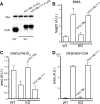
Figure 4. Enzyme activity of CLYBL is required to support mitochondrial B12 in brown adipocytes
(A) Western blot of FLAG-tagged wild type human CLYBL and catalytic dead mutant CLYBLD320A in Clybl KO cells. (B–D) Methylmalonate (MMA) accumulation (in B), coenzyme B12 inhibition (in C), and citramalyl-CoA accumulation (in D) in Clybl KO cellular extract cannot be rescued by catalytically dead mutant of CLYBL.
Figure 5
Figure 5. Itaconyl-CoA directly inactivates coenzyme B12 through MUT
(A) Cellular methylmalonate (MMA) is accumulated in control brown adipocytes treated with 2 mM itaconate. (B) Coenzyme B12 is reduced in control brown adipocytes treated with 2 mM itaconate. (C) Western blot of Clybl, Mut in double KO cells. (D–E) Loss of Mut suppresses coenzyme B12 deficiency in Clybl KO cells (in D), despite similarly increased MMA level (in E), suggesting that the inhibition mechanism is via MUT activity. (F–G) Itaconyl-CoA inactivates human MUT enzyme. Purified human MUT enzyme was preloaded with coenzyme B12 AdoCbl (with an absorbance maximum at 529 nm). UV-visible spectra were recorded before (red), immediately after (gray), and 20 min after (black) addition of either substrate M-CoA (in F) or itaconyl-CoA (in G). Unlike M-CoA, which elicited only minor spectral changes, itaconyl-CoA triggered a large blue shift in the absorbance maximum to 466 nm, indicating formation of inactive cob(II)alamin.
Figure 6
Figure 6. The immunomodulatory metabolite itaconate poisons vitamin B12 in various cell types
(A–B) Coenzyme B12 is reduced by 2 mM itaconate treatment in HEK293T cell (in A), and in human B-lymphocytes (in B). Itaconyl-CoA is undetected in untreated cells, but dramatically increased in treated cells. (C) Coenzyme B12 is ablated in macrophages upon 10 ng/ml LPS stimulation for 6 hr. Itaconyl-CoA is dramatically increased upon stimulation.
Figure 7
Figure 7. Relationship between CLYBL, itaconate and B12
CLYBL functions as a citramalyl-CoA lyase, and its loss leads to an accumulation of upstream metabolites. Itaconyl-CoA serving as a substrate analog directly inhibits the MUT enzyme and rapidly inactivates the coenzyme B12.

Comment in

References

    1. Adams PD, Afonine PV, Bunkoczi G, Chen VB, Davis IW, Echols N, Headd JJ, Hung LW, Kapral GJ, Grosse-Kunstleve RW, et al. PHENIX: a comprehensive Python-based system for macromolecular structure solution. Acta Crystallogr. D Biol. Crystallogr. 2010;66:213–221. - PMC - PubMed
    1. Adler J, Wang SF, Lardy HA. The metabolism of itaconic acid by liver mitochondria. J. Biol. Chem. 1957;229:865–879. - PubMed
    1. Banerjee R, Gherasim C, Padovani D. The tinker, tailor, soldier in intracellular B12 trafficking. Curr. Opin. Chem. Biol. 2009;13:484–491. - PMC - PubMed
    1. Banerjee R, Ragsdale SW. The many faces of vitamin B12: catalysis by cobalamin-dependent enzymes. Annu. Rev. Biochem. 2003;72:209–247. - PubMed
    1. Bracken CD, Neighbor AM, Lamlenn KK, Thomas GC, Schubert HL, Whitby FG, Howard BR. Crystal structures of a halophilic archaeal malate synthase from Haloferax volcanii and comparisons with isoforms A and G. BMC Struct. Biol. 2011;11:23. - PMC - PubMed