. 2018 Jan;299(Pt A):86-96.
doi: 10.1016/j.expneurol.2017.10.013. Epub 2017 Oct 19.

Photobiomodulation therapy promotes neurogenesis by improving post-stroke local microenvironment and stimulating neuroprogenitor cells

Affiliations

Photobiomodulation therapy promotes neurogenesis by improving post-stroke local microenvironment and stimulating neuroprogenitor cells

Luodan Yang et al. Exp Neurol. 2018 Jan.

Abstract

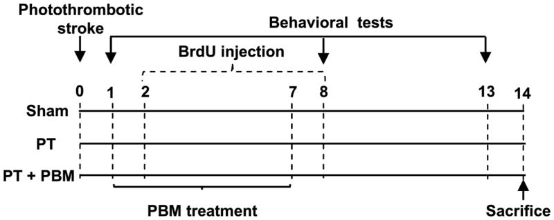
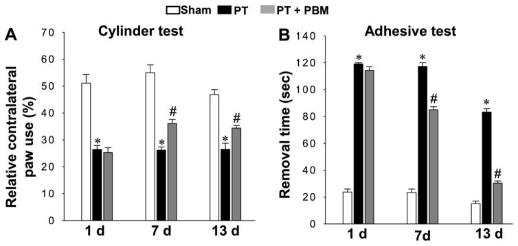
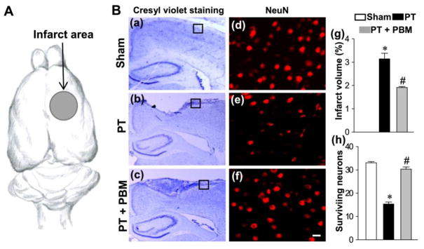
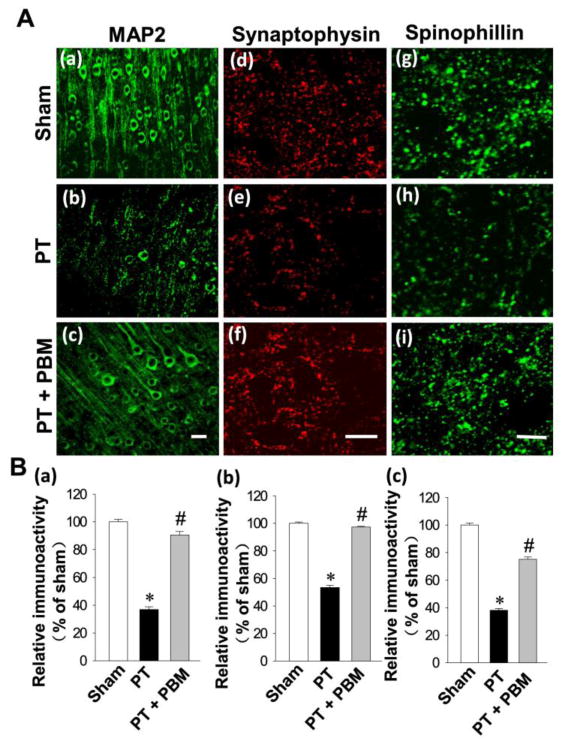
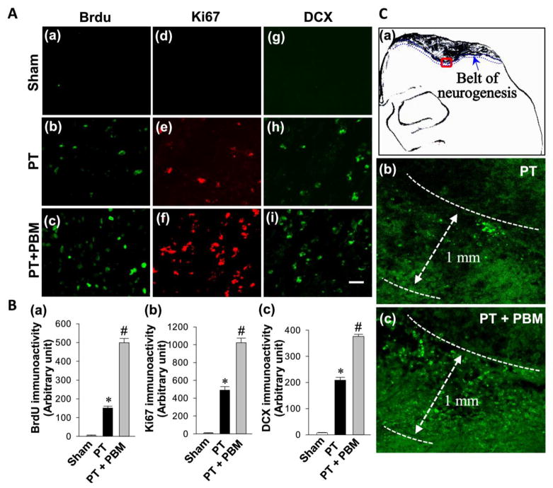
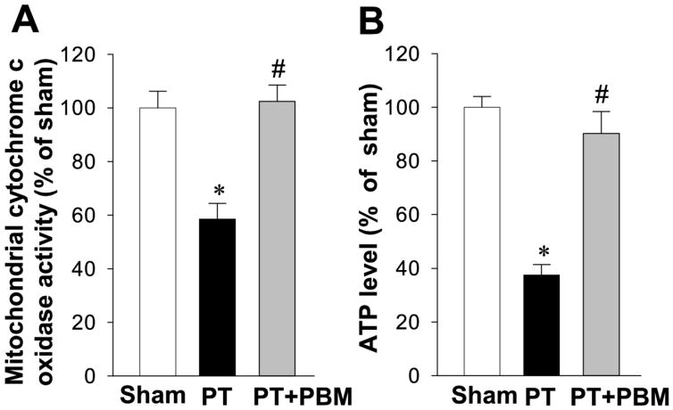
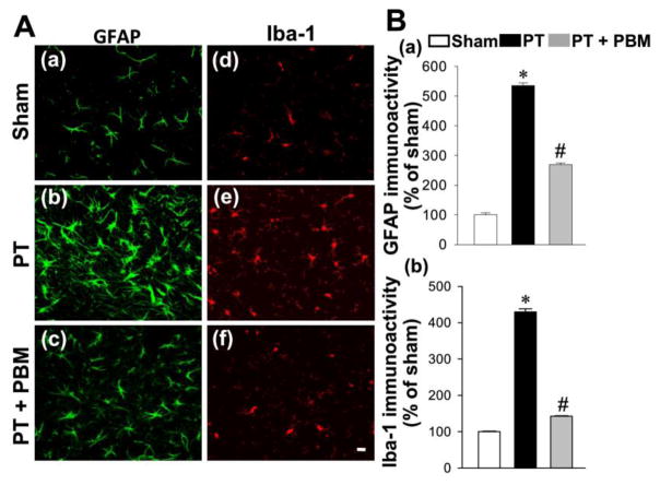
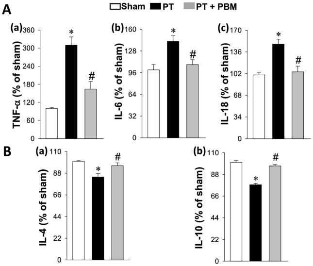
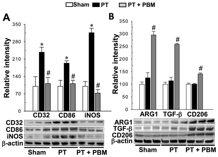
Recent work has indicated that photobiomodulation (PBM) may beneficially alter the pathological status of several neurological disorders, although the mechanism currently remains unclear. The current study was designed to investigate the beneficial effect of PBM on behavioral deficits and neurogenesis in a photothrombotic (PT) model of ischemic stroke in rats. From day 1 to day 7 after the establishment of PT model, 2-minute daily PBM (CW, 808nm, 350mW/cm2, total 294J at scalp level) was applied on the infarct injury area (1.8mm anterior to the bregma and 2.5mm lateral from the midline). Rats received intraperitoneal injections of 5-bromodeoxyuridine (BrdU) twice daily (50mg/kg) from day 2 to 8 post-stoke, and samples were collected at day 14. We demonstrated that PBM significantly attenuated behavioral deficits and infarct volume induced by PT stroke. Further investigation displayed that PBM remarkably enhanced neurogenesis and synaptogenesis, as evidenced by immunostaining of BrdU, Ki67, DCX, MAP2, spinophilin, and synaptophysin. Mechanistic studies suggested beneficial effects of PBM were accompanied by robust suppression of reactive gliosis and the production of pro-inflammatory cytokines. On the contrary, the release of anti-inflammatory cytokines, cytochrome c oxidase activity and ATP production in peri-infarct regions were elevated following PBM treatment. Intriguingly, PBM could effectively switch an M1 microglial phenotype to an anti-inflammatory M2 phenotype. Our novel findings indicated that PBM is capable of promoting neurogenesis after ischemic stroke. The underlying mechanisms may rely on: 1) promotion of proliferation and differentiation of internal neuroprogenitor cells in the peri-infarct zone; 2) improvement of the neuronal microenvironment by altering inflammatory status and promoting mitochondrial function. These findings provide strong support for the promising therapeutic effect of PBM on neuronal repair following ischemic stroke.

Keywords: Inflammation; Ischemic stroke; Mitochondrial function; Neurogenesis; Photobiomodulation.


Conflict of interest statement

Statement of interest

The authors declare that there is no conflict of interest in the current study.

Figures

Fig. 1
Fig. 1. Schematic diagram of the experimental protocols
PT model was induced on day 0, and on days 1 through 7, rats were treated with PBM (2-minute daily). BrdU (50 mg/kg, ip) was injected two times daily on days 2 through 8. Behavioral deficits were evaluated on days 1, 7 and 13. Rats were sacrificed on day 14, and whole brains were extracted and prepared for further analysis.
Fig. 2
Fig. 2. Effect of PBM on behavioral deficits in PT stroke rats
(A) Results of the cylinder test. The percentage of relative contralateral paw use was used to evaluate rats’ spontaneous forelimb use. (B) Results of the adhesive removal test. The time spent on removing a small rectangular adhesive strip was recorded and used for measuring stimulus-directed movement after PT. Data are expressed as mean ± SE (n = 8–10). *P < 0.05 versus sham group, #P < 0.05 versus PT.
Fig. 3
Fig. 3. Effect of PBM on the cortical infarct size and the number of surviving neurons
(A) The area illuminated by cold fiber light was pointed out by an arrow presenting somatosensory cortex. (B) Representative images of cresyl violet staining (a–c) and confocal images of NeuN (d–f). The surviving neuros were counted from the locations in peri-infarct zone indicated by the boxes in (a–c). The surviving neurons (h) were counted from images like those in (d–f) taken from locations like those indicated by the boxes in (a–c). Infarct volume was calculated and expressed as the percentage of the total volume of the contralateral hemisphere per group in (g). The count of surviving neurons was analyzed in (h). All data are expressed as mean ± SE (n = 4–5). *P < 0.05 versus sham, #P < 0.05 versus PT. Scale bar =10 μm.
Fig. 4
Fig. 4. Effect of PBM on PT stroke-induced neuronal injury in the infarct cortical region 14 days after PT stroke
(A) Typical staining of MAP2 in healthy control animals (a), animals that underwent PT stroke without (b) or with PBM treatment (c). Representative confocal microscopy images of synaptophysin (d–f) and spinophilin (g–i) were taken from the infarct cortical region 14 days after PT stroke. (B) The fluorescent intensity of MAP2 (a), synaptophysin (b) and spinophilin (c) were quantified using Image J analysis software and expressed as percentage changes versus the respective control group. (C) The region in which neurogenesis is illustrated (a) and representative images from PT (b) and PBM treated rats (c) are displayed. All data are expressed as mean ± SE (n = 4–5). *P < 0.05 versus sham, #P < 0.05 versus PT. Scale bar =10 μm.
Fig. 5
Fig. 5. LLI improves neurogenesis in the peri-infarct cortical region on day 14 after PT stroke
(A) Representative confocal microscopy images of BrdU (a–c), Ki67 (d–f) and DCX (g–i) were taken from the cortical peri-infarct region on day 14 after PT stroke. (B) The immunoactivity associated with BrdU, Ki67 and DCX in each group were further quantified and shown in arbitrary unit (a, b and c respectively). (C) The increased neurogenesis was restricted to a regions (around 1.00 mm wide belt) beneath the infarct zone surface with the representative confocal microscopy of Brdu staining (a–c). All data are expressed as mean ± SE (n = 4–5). *P < 0.05 versus sham, #P < 0.05 versus PT. Scale bar = 20 μm.
Fig. 6
Fig. 6. Effect of PBM on the activity of mitochondrial cytochrome c oxidase and the production of ATP in the cortical peri-infarct region
Mitochondrial cytochrome c oxidase activity (A) and ATP production (B) in total protein samples were measured. The results of PT stroke group and PBM group are quantified as percentage changes versus sham. Values are expressed as mean±SE (n=5). *P<0.05 versus sham, #P<0.05 versus PT group.
Fig. 7
Fig. 7. PBM inhibits PT stroke-induced glial activation
(A) Representative confocal microscopy of GFAP (a–c) and Iba-1 (d–f) staining taken from the peri-infarct cortical region. (B) The fluorescent intensity of GFAP (a) and Iba-1(b) was quantified and expressed as percentage changes versus the respective control group. Values are expressed as mean ± SE (n=4–5). *P<0.05 versus sham, #P<0.05 versus PT group.
Fig. 8
Fig. 8. Effect of PB< on inflammatory cytokines
(A) The levels of pro-inflammatory cytokines, TNF-a, IL-6 and IL-18 were measured and shown in (a–c). (B) The levels of anti-inflammatory cytokines, IL-4 and IL-10 were also were measured by ELISA assay (a–b). All data are expressed as mean ± SE (n = 4–5). *P < 0.05 versus sham, #P < 0.05 versus PT.
Fig. 9
Fig. 9. PBM shifts microglial polarization from M1 to M2 phenotype
(A) Results of western blot analysis and quantitative analyses for M1 markers, CD32, CD86 and iNOS. (B) indicates the effect of LLI on M2 markers, ARG1, TGF-β and CD 206. All data are expressed as mean ± SE (n = 4–5). *P < 0.05 versus sham, #P < 0.05 versus PT.

References

    1. Ahmed ME, Tucker D, Dong Y, Lu Y, Zhao N, Wang R, Zhang Q. Methylene Blue promotes cortical neurogenesis and ameliorates behavioral deficit after photothrombotic stroke in rats. Neuroscience. 2016;336:39–48. - PMC - PubMed
    1. Alcantara CC, Gigo-Benato D, Salvini TF, Oliveira AL, Anders JJ, Russo TL. Effect of low-level laser therapy (LLLT) on acute neural recovery and inflammation-related gene expression after crush injury in rat sciatic nerve. Lasers in surgery and medicine. 2013;45:246–252. - PubMed
    1. Ando T, Xuan W, Xu T, Dai T, Sharma SK, Kharkwal GB, Huang YY, Wu Q, Whalen MJ, Sato S, Obara M, Hamblin MR. Comparison of therapeutic effects between pulsed and continuous wave 810-nm wavelength laser irradiation for traumatic brain injury in mice. PloS one. 2011;6:e26212. - PMC - PubMed
    1. Andrabi SS, Parvez S, Tabassum H. Progesterone induces neuroprotection following reperfusion-promoted mitochondrial dysfunction after focal cerebral ischemia in rats. Disease models & mechanisms. 2017;10:787–796. - PMC - PubMed
    1. Arvidsson A, Collin T, Kirik D, Kokaia Z, Lindvall O. Neuronal replacement from endogenous precursors in the adult brain after stroke. Nature medicine. 2002;8:963–970. - PubMed

MeSH terms