. 2018 Jan;299(Pt A):97-108.
doi: 10.1016/j.expneurol.2017.10.014. Epub 2017 Oct 19.

Apolipoprotein E as a novel therapeutic neuroprotection target after traumatic spinal cord injury

Affiliations

Apolipoprotein E as a novel therapeutic neuroprotection target after traumatic spinal cord injury

Xiaoxin Cheng et al. Exp Neurol. 2018 Jan.

Abstract

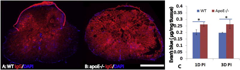
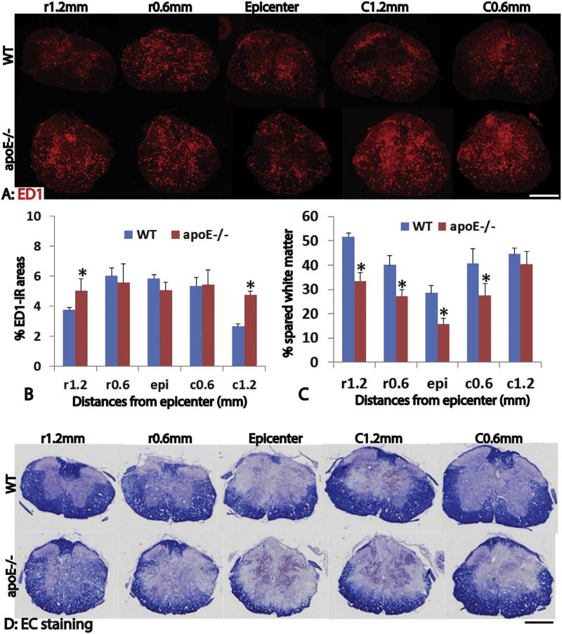
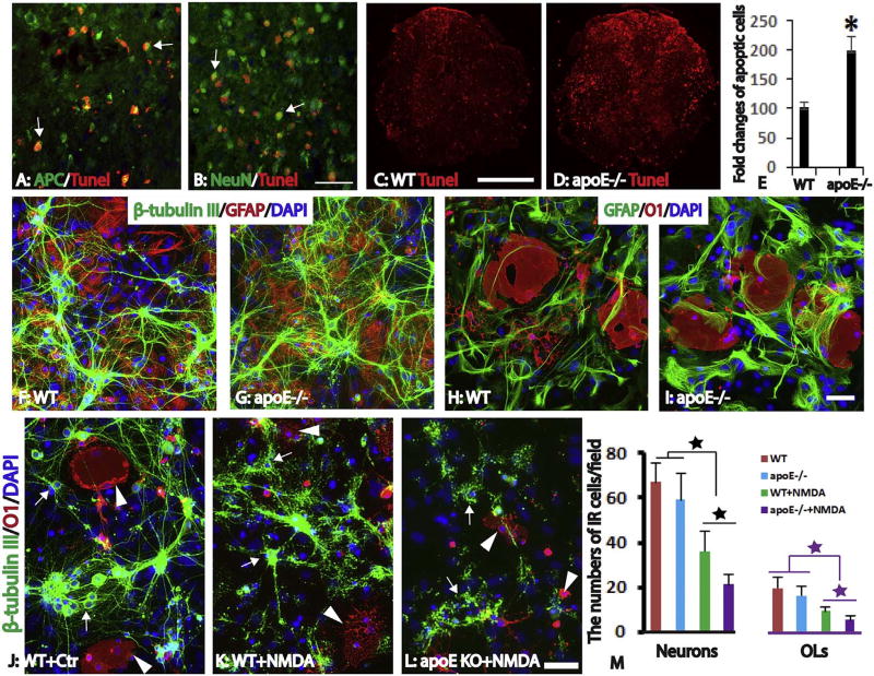
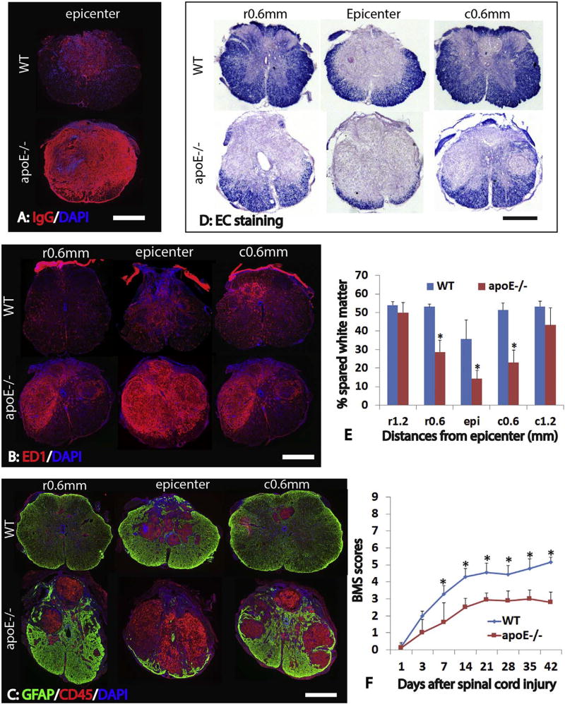
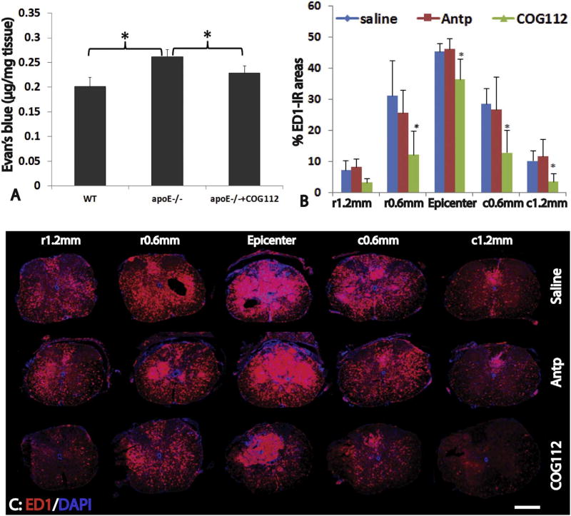
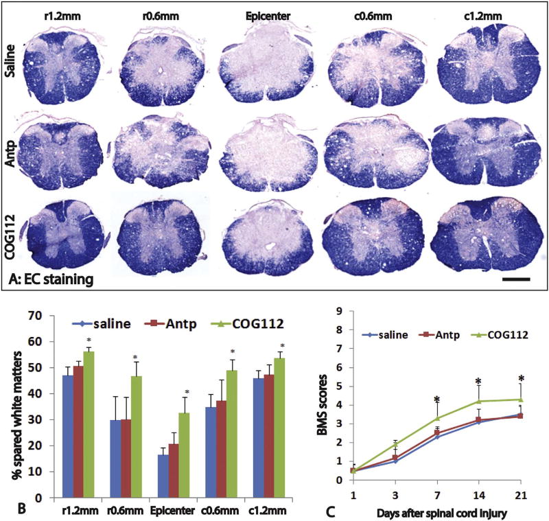
Apolipoprotein E (apoE), a plasma lipoprotein well known for its important role in lipid and cholesterol metabolism, has also been implicated in many neurological diseases. In this study, we examined the effect of apoE on the pathophysiology of traumatic spinal cord injury (SCI). ApoE-deficient mutant (apoE-/-) and wild-type mice received a T9 moderate contusion SCI and were evaluated using histological and behavioral analyses after injury. At 3days after injury, the permeability of spinal cord-blood-barrier, measured by extravasation of Evans blue dye, was significantly increased in apoE-/- mice compared to wild type. The inflammation and spared white matter was also significantly increased and decreased, respectively, in apoE-/- mice compared to the wild type ones. The apoptosis of both neurons and oligodendrocytes was also significantly increased in apoE-/- mice. At 42days after injury, the inflammation was still robust in the injured spinal cord in apoE-/- but not wild type mice. CD45+ leukocytes from peripheral blood persisted in the injured spinal cord of apoE-/- mice. The spared white matter was significantly decreased in apoE-/- mice compared to wild type ones. Locomotor function was significantly decreased in apoE-/- mice compared to wild type ones from week 1 to week 8 after contusion. Treatment of exogenous apoE mimetic peptides partially restored the permeability of spinal cord-blood-barrier in apoE-/- mice after SCI. Importantly, the exogenous apoE peptides decreased inflammation, increased spared white matter and promoted locomotor recovery in apoE-/- mice after SCI. Our results indicate that endogenous apoE plays important roles in maintaining the spinal cord-blood-barrier and decreasing inflammation and spinal cord tissue loss after SCI, suggesting its important neuroprotective function after SCI. Our results further suggest that exogenous apoE mimetic peptides could be a novel and promising neuroprotective reagent for SCI.

Keywords: Apolipoprotein E; Neuroprotection; Spinal cord injury; Spinal cord-blood-barrier; Treatment.


Figures

Fig. 1
Fig. 1
Increased permeability of blood-spinal cord-barrier (BSCB) in apoE deficient mice after SCI. BSCB permeability was evaluated by the extravasation of IgG using immunohistochemistry (A, B). Compared to wild type B6 mice (A), the extravasation of circulating IgG in the injury epicenter was significantly increased in apoE−/− mice at 3 days after SCI (B). Quantification of BSCB permeability using Evan Blue technique also showed that the amount of EB in the injured spinal cord was significantly increased in apoE−/− mice compared to wild type B6 mice at 1 day and 3 days after SCI. Scale bar = 500 µm in A, B. Data in C represent the mean ± SD, N = 4, stars represent p < 0.05.
Fig. 2
Fig. 2
Increased inflammation and neural tissue loss in apoE deficient mice after acute SCI. Inflammation was evaluated by immunohistochemistry of ED1, a marker for macrophage and activated microglia. Three days after SCI, inflammation was significantly increased in apoE deficient mice, especially at 1.2 mm from injury epicenter both rostrally and caudally (A, B). Importantly, the spared white matter shown by EC staining was significantly decreased in apoE deficient mice compared to wild type mice (C, D). Scale bar = 500 µm in A and D. Data in B and C represent the mean ± SD, N = 4, stars represent p < 0.05.
Fig. 3
Fig. 3
Increased neuron and oligodendrocyte loss in apoE−/− mice in vivo and in vitro. Robust apoptosis shown by TUNEL staining was observed in the injured spinal cord at 3 days after SCI (A–D). At the injury epicenter, most TUNNEL + apoptotic cells are oligodendrocytes (A, arrows) and neurons (B, arrows). The number of apoptotic cells in the injured epicenter were significantly increased in apoE−/− mice (C–D) and doubled in apoE−/− mice compared to WT mice (E). To further examine the effects of endogenous apoE in the survival of neurons and oligodendrocytes, neurons, astrocytes and oligodendrocytes were isolated from postnatal day 3 spinal cord of WT or apoE−/− mice and co-cultured for 7 days (F–I). In the control culture condition, the morphology and number of neurons (F, G) or oligodendrocytes (H, I) was not significantly different between WT or apoE−/− mice. Treatment with NMDA resulted in the death of neurons and oligodendrocytes derived from WT (K) and apoE−/− (L) mice. The surviving neurons (arrows) and oligodendrocytes (arrowheads) from WT or apoE−/− mice were significantly decreased after NMDA treatment compared to its respective un-treated counterpart (Fig. J–M). Importantly, the numbers of survived neurons or oligodendrocytes from apoE−/− spinal cord were further decreased to only half of those from WT mice spinal cord after NMDA treatment (M). Scale bar = 50 µm in A, B, 500 µm in C, D, 50 µm in F–I and J–L. Data in E and M represent the mean ± SD, N = 4 (E) and 6 (M), stars represent p < 0.05.
Fig. 4
Fig. 4
Increased anatomical deficiency in apoE−/− mice after SCI. At 6 weeks after SCI, the extravasation of circulating IgG was significantly increased in the injured spinal cord epicenter in apoE−/− mice compared to WT ones (A). The numbers of ED1+ macrophages and active microglial cells were significantly increased at the injured epicenter as well as at 0.6 mm from epicenter rostrally and caudally in apoE−/− mice compared to WT ones (B). Similarly, the numbers of CD45+ lymphocytes were significantly increased in these injury levels in apoE−/− mice (C). Conversely, the spared white matter was significantly decreased in apoE−/− mice at the injured epicenter as well as 0.6 mm rostrally and caudally from the epicenter (D, E). Importantly, locomotion function assessed by Basso-Mouse-Score (BMS) was significantly worsen in apoE−/− mice starting at 1 week after SCI (F). Scale bar = 500 µm in A, B, and C. Data in D and E represent the mean ± SD, N = 6 (D) and 10 (E), stars represent p < 0.05.
Fig. 5
Fig. 5
Treatment of exogenous apoE mimetic peptides decreases the BSCB permeability and inflammation in apoE−/− mice after SCI. At 3 days after SCI, permeability of BSCB shown by the extravasation of Evan Blue was significantly increased in apoE−/− mice compared to WT (A). However, the deteriorated permeability of BSCB in apoE−/− mice was significantly attenuated after treatment with exogenous apoE mimic peptides following SCI (A). Importantly, treatment of exogenous apoE mimic peptides also significantly decreased inflammation in apoE−/− mice at 21 days after SCI (B, C). Scale bar = 500 µm in C. Data in A and B represent the mean ± SD, N = 4 (D) and 6 (E), stars represent p < 0.05.
Fig. 6
Fig. 6
Treatment of exogenous apoE mimetic peptides improves the anatomical and functional recovery in apoE−/− mice after SCI. The spared white matter was significantly increased in increased in apoE−/− mice treated by exogenous mimic apoE peptides compared to ones treated by saline or control Antp peptides (A, B). Importantly, locomotion function was also significantly increased in apoE−/− mice treated by exogenous mimic apoE peptides compared to ones treated by saline or control Antp peptides from 1 week to 3 weeks post-injury, the longest time for treatment (C). Scale bar = 500 µm in A. Data in B and C represent the mean ± SD, N = 6 (B) and 10 (C), stars represent p < 0.05.

References

    1. Alberts MJ, Graffagnino C, McClenny C, DeLong D, Strittmatter W, Saunders AM, Roses AD. ApoE genotype and survival from intracerebral haemorrhage. Lancet. 1995;346:575. - PubMed
    1. Aono M, Bennett ER, Kim KS, Lynch JR, Myers J, Pearlstein RD, Warner DS, Laskowitz DT. Protective effect of apolipoprotein E-mimetic peptides on N-methyl-d-aspartate excitotoxicity in primary rat neuronal-glial cell cultures. Neuroscience. 2003;116:437–445. - PubMed
    1. Badaut J, Copin JC, Fukuda AM, Gasche Y, Schaller K, da Silva RF. Increase of arginase activity in old apolipoprotein-E deficient mice under Western diet associated with changes in neurovascular unit. J. Neuroinflammation. 2012;9:132. - PMC - PubMed
    1. Bart RD, Sheng H, Laskowitz DT, Pearlstein RD, Warner DS. Regional CBF in apolipoprotein E-deficient and wild type mice during focal cerebral ischemia. Neuroreport. 1998;9:2615–2620. - PubMed
    1. Basso DM, Fisher LC, Anderson AJ, Jakeman LB, McTigue DM, Popovich PG. Basso Mouse Scale for locomotion detects differences in recovery after spinal cord injury in five common mouse strains. J. Neurotrauma. 2006;23:635–659. - PubMed

Publication types

MeSH terms