. 2018 Feb 16;430(4):479-490.
doi: 10.1016/j.jmb.2017.10.012. Epub 2017 Oct 19.

Two Disease-Causing SNAP-25B Mutations Selectively Impair SNARE C-terminal Assembly

Affiliations

Two Disease-Causing SNAP-25B Mutations Selectively Impair SNARE C-terminal Assembly

Aleksander A Rebane et al. J Mol Biol. .

Abstract

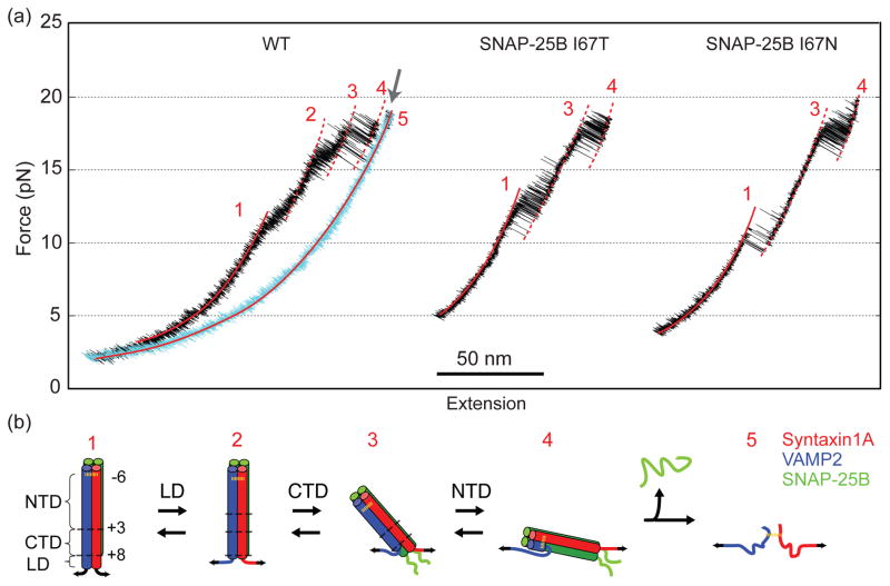
Synaptic exocytosis relies on assembly of three soluble N-ethylmaleimide-sensitive factor attachment receptor (SNARE) proteins into a parallel four-helix bundle to drive membrane fusion. SNARE assembly occurs by stepwise zippering of the vesicle-associated SNARE (v-SNARE) onto a binary SNARE complex on the target plasma membrane (t-SNARE). Zippering begins with slow N-terminal association followed by rapid C-terminal zippering, which serves as a power stroke to drive membrane fusion. SNARE mutations have been associated with numerous diseases, especially neurological disorders. It remains unclear how these mutations affect SNARE zippering, partly due to difficulties to quantify the energetics and kinetics of SNARE assembly. Here, we used single-molecule optical tweezers to measure the assembly energy and kinetics of SNARE complexes containing single mutations I67T/N in neuronal SNARE synaptosomal-associated protein of 25kDa (SNAP-25B), which disrupt neurotransmitter release and have been implicated in neurological disorders. We found that both mutations significantly reduced the energy of C-terminal zippering by ~10 kBT, but did not affect N-terminal assembly. In addition, we observed that both mutations lead to unfolding of the C-terminal region in the t-SNARE complex. Our findings suggest that both SNAP-25B mutations impair synaptic exocytosis by destabilizing SNARE assembly, rather than stabilizing SNARE assembly as previously proposed. Therefore, our measurements provide insights into the molecular mechanism of the disease caused by SNARE mutations.

Keywords: SNARE assembly; membrane fusion; neuropathy; optical tweezers; protein folding.


Figures

FIGURE 1
FIGURE 1
SNARE complex and experimental setup. The ternary SNARE complex forms a parallel four-helix bundle that is stabilized by inward-facing residues in layers −7 to +8. Engineered cysteines at the -6 layer create a disulfide bridge between syntaxin and VAMP2 to facilitate SNARE re-assembly. SNARE assembly occurs by sequential folding of the NTD, the CTD, and the LD. The N-terminal Habc domain in syntaxin recruits other proteins to regulate SNARE assembly [45, 46], but minimally affects ternary SNARE assembly in the absence of these regulatory proteins in our assay [13]. Disease-causing mutations SNAP-25B I67T and I67N are located in the +4 layer.
FIGURE 2
FIGURE 2
SNAP-25B mutations destabilize SNARE CTD. (a) FECs obtained by pulling (black) or relaxing (cyan) single SNARE complexes. Different SNARE folding states are marked by red numbers of states depicted in b. These states are derived from continuous regions in the FECs (red solid curves) or regions with discrete but distinct extensions (red dashed lines) based on the worm-like chain model [32]. (b) Diagrams of different SNARE folding states. The folding states of the WT SNARE complex include the fully assembled SNARE state (state 1), the LD-unfolded four-helix bundle state (2), the partially zippered state (3), the unzipped state (4), and the fully unfolded state (5). Folding of both mutant SNARE complexes I67T and I67N bypasses the state 2.
FIGURE 3
FIGURE 3
Representative extension-time trajectories containing the LD/CTD transition for I67T and I67N or the CTD transition for WT. The mean force F was kept constant for each trajectory by fixing the distance between two optical traps. Red traces represent idealized state trajectories as determined by HMM. Double-Gaussian fits (green curves) of the extension probability density distributions (gray dots) reveal transitions between the two discrete states indicated by their corresponding state numbers (Fig. 2b). All extension traces share the same length and time scale bars, except for the trace at the bottom, which has a different time scale bar for a close-up view.
FIGURE 4
FIGURE 4
Zippering energy and kinetics of WT and mutant SNARE complexes. (a, b) Force-dependent unfolding probabilities (top panel) and transition rates (bottom panel) for CTD and LD/CTD transitions (a) or NTD transitions (b). Symbols denote measurements from extension-time trajectories for CTD transition in WT (black circles) and LD/CTD transition in I67T (red diamonds) or I67N (blue squares). Folding and unfolding rates are shown as hollow and solid symbols, respectively. Curves represent fitting results with a non-linear two-state model (Materials and Methods). Gray arrows mark transition rates at the corresponding equilibrium forces. (c) Comparison of NTD (gray) and LD/CTD (red) zippering energies. (d) Simplified energy landscape of SNARE zippering at zero force. The abscissa denotes the VAMP2 residue to which the SNARE complex is structured starting from the cross-linking site at -6 layer (residue 36). The regions corresponding to NTD, CTD, and LD are marked at the top of the graph. The derived stable and transition states are denoted by solid and hollow symbols, respectively. Solid lines denote an arbitrary interpolation between the calculated states to guide the eye.
FIGURE 5
FIGURE 5
Conformations and dynamics of WT and mutant t-SNARE complexes. (a) The correctly folded t-SNARE complex (state iii) is prepared by completely unfolding a ternary SNARE complex (state i) in situ at high force and subsequent refolding the remaining t-SNAREs (state ii). Note that SNAP-25B contains an N-terminal SNARE domain (SN1) and a C-terminal SNARE domain (SN2) connected by a disordered linker. The t-SNARE complex is pulled from the C-termini of syntaxin and SN1. (b) FECs obtained by pulling t-SNARE complexes in ternary SNARE complexes (black) and then relaxing the t-SNARE complexes alone (cyan). Green arrows indicate CTD transitions in ternary SNARE complexes. (c) Representative extension-time trajectories for the t-SNARE folding/unfolding transition near equilibrium force. Double-Gaussian fits (green curves) of the extension probability density distributions (gray dots) confirm the two-state nature of the transition. Red traces represent idealized state trajectories.
FIGURE 6
FIGURE 6
Folding energies, kinetics, and conformations of t-SNARE complexes. (a) Force-dependent unfolding probabilities (top panel) and transition rates (bottom panel) of the t-SNARE complex. Symbols denote experimental measurements for WT (black circles), I67T (red diamonds), and I67N (blue squares). Folding and unfolding rates are shown as hollow and solid symbols, respectively. Best-fits with a two-state model are shown as curves. (b) Comparison of t-SNARE folding energies between WT and mutant complexes. (c) Simplified folding energy landscapes for t-SNARE complexes. The abscissa denotes the syntaxin residue to which the t-SNARE complex is structured starting from the cross-linking site at -8 layer (residue 199). Locations of corresponding hydrophobic and ionic layers are marked on top of the graph. The derived stable and transition states are shown as solid and hollow symbols, respectively, for WT (black), I67T (red), and I67N (blue).

References

    1. Sudhof TC, Rothman JE. Membrane fusion: grappling with SNARE and SM proteins. Science. 2009;323:474–7. - PMC - PubMed
    1. Weber T, Zemelman BV, McNew JA, Westermann B, Gmachl M, Parlati F, et al. SNAREpins: Minimal machinery for membrane fusion. Cell. 1998;92:759–72. - PubMed
    1. Sollner T, Whiteheart SW, Brunner M, Erdjument-Bromage H, Geromanos S, Tempst P, et al. SNAP receptors implicated in vesicle targeting and fusion. Nature. 1993;362:318–24. - PubMed
    1. Bennett MK, Calakos N, Scheller RH. Syntaxin - a synaptic protein implicated in docking of synaptic vesicles at presynaptic active zones. Science. 1992;257:255–9. - PubMed
    1. Trimble WS, Cowan DM, Scheller RH. VAMP-1 - a synaptic vesicle-associated integral membrane-protein. Proc Natl Acad Sci USA. 1988;85:4538–42. - PMC - PubMed

Publication types

Substances

LinkOut - more resources