. 2017 Nov 7;25(11):1708-1718.e5.
doi: 10.1016/j.str.2017.09.010. Epub 2017 Oct 19.

Solution Structure and Membrane Interaction of the Cytoplasmic Tail of HIV-1 gp41 Protein

Affiliations

Solution Structure and Membrane Interaction of the Cytoplasmic Tail of HIV-1 gp41 Protein

R Elliot Murphy et al. Structure. .

Abstract

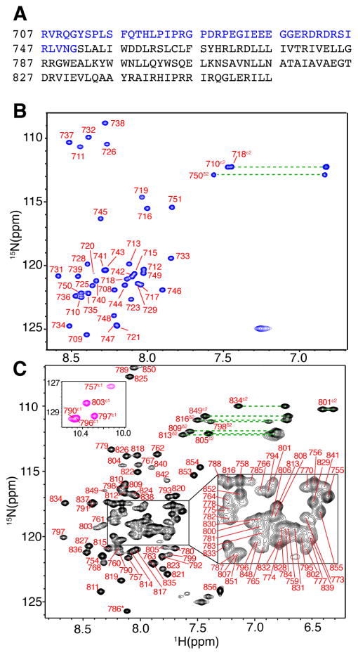
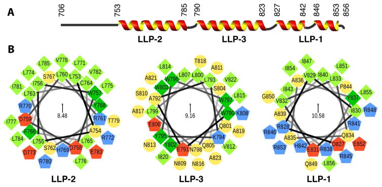
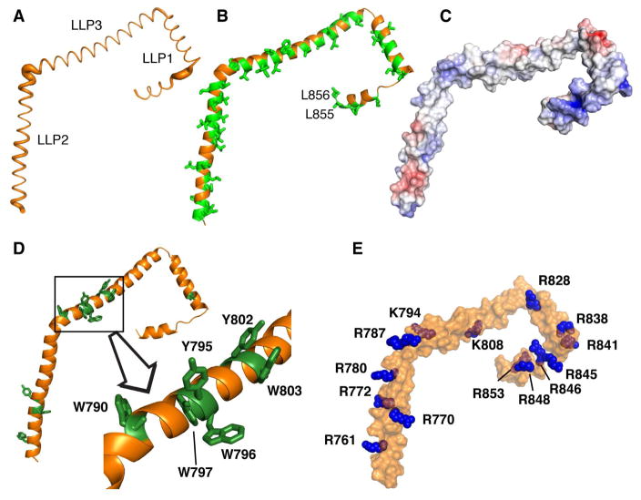
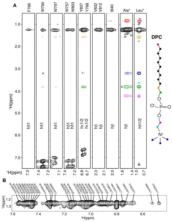
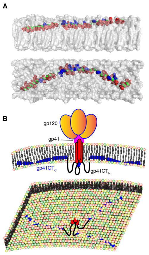
The cytoplasmic tail of gp41 (gp41CT) remains the last HIV-1 domain with an unknown structure. It plays important roles in HIV-1 replication such as mediating envelope (Env) intracellular trafficking and incorporation into assembling virions, mechanisms of which are poorly understood. Here, we present the solution structure of gp41CT in a micellar environment and characterize its interaction with the membrane. We show that the N-terminal 45 residues are unstructured and not associated with the membrane. However, the C-terminal 105 residues form three membrane-bound amphipathic α helices with distinctive structural features such as variable degree of membrane penetration, hydrophobic and basic surfaces, clusters of aromatic residues, and a network of cation-π interactions. This work fills a major gap by providing the structure of the last segment of HIV-1 Env, which will provide insights into the mechanisms of Gag-mediated Env incorporation as well as the overall Env mobility and conformation on the virion surface.

Keywords: Gag polyprotein; HIV-1; NMR; bicelles; cytoplasmic tail; envelope protein; gp41; matrix protein; membrane; micelles.


Figures

Figure 1
Figure 1. NMR Spectra of the Two Independent gp41CT Domains
(A) Amino acid sequence of the HIV-1 gp41CT protein (pNL4-3 isolate). Sequences of gp41CTN and gp41CTC are highlighted in blue and black, respectively. (B) 2D 1H-15N HSQC spectrum obtained for gp41CTN (200 μM) at 32 °C. (C) 2D 1H-15N HSQC spectrum obtained for gp41CTC (300 μM) at 50 °C. Tryptophan side chain signals are colored in magenta (inset). Dashed lines indicate side chain amide signals.
Figure 2
Figure 2. Secondary Structure and Helical Wheel Diagrams of the gp41CTC Protein
(A) Secondary structure representation of the gp41CT protein based on the NMR data. (B) Helical wheel diagrams of the LLP2, LLP3, and LLP1 motifs. Amino acid sequences are plotted clockwise. Hydrophobic and aromatic residues are represented by light and dark green squares, respectively. Polar residues are shown as yellow circles, while positively and negatively charged residues as blue and red pentagons, respectively. The helical wheels are oriented so that their hydrophobic moments (indicated in the centers) point upwards. Helical wheels were generated via a modified script obtained from http://rzlab.ucr.edu/scripts/wheel/.
Figure 3
Figure 3. Structural Features of the gp41CTC Protein
(A) The Cα positional RMSDs between the 20 low-energy structures of gp41CTC are represented as a sausage plot of the representative gp41CTC structure (line thickness is proportional to the RMSD values). The RMSDs from an average structure were calculated after aligning each α-helical segment separately. For residues located at the overlapping boundaries of individual segments, an average of the two RMSD values was calculated. Bending between the helical fragments is variable in the 20 calculated structures. (B) Cartoon representation of the gp41CTC protein showing the extensive hydrophobic interface formed by Leu, Ile, Val, Ala, Trp and Phe residues (green sticks). (C) Surface representation of the gp41CTC colored according to electrostatic surface potential. A basic patch is formed in the arginine-rich LLP1 motif. (D) Cartoon representation of the gp41CTC protein showing Tyr and Trp residues as green sticks. Notice the cluster of six aromatic residues at the beginning of LLP3. The majority of aromatic residues are deeply buried in the interior of DPC micelle. (E) Surface representation of the gp41CTC protein showing Arg and Lys residues (blue spheres).
Figure 4
Figure 4. Intermolecular Contacts Between gp41CTC and Micelles
(A) 3D 13C-half-filtered/13C-edited NOESY spectrum for 13C-labeled gp41CTC showing intermolecular NOEs between DPC and gp41CTC residues. Colored intermolecular NOE cross-peaks correspond to colored atoms on the DPC structure. (B) A selected region of 2D 1H-1H NOESY spectrum for gp41CTC showing NOEs between side chains of aromatic residues and DPC acyl chain methylene groups.
Figure 5
Figure 5. Membrane Interaction of gp41CTC and Overall Env Organization on the Virion Surface
(A) A model of gp41CTC bound to a membrane bilayer constructed based on the NMR data using the representative structure of gp41CTC with only minor modifications of the dihedral angles in the hinge regions to create an extended molecule. Length of the extended gp41CTC domain shown here is 160 Å. Top and bottom panels show side and top views of the protein, respectively. Residues indicated as red spheres interact extensively with the interior of the membrane while those in blue are mostly exposed and interact with the polar head. Membrane bilayer was generated in VMD membrane builder plug-in (Humphrey et al., 1996). (B) Top panel: A model depicting the gp120 and gp41 proteins on the surface of HIV-1 particles. The gp41CTC domain is penetrating deeply in the inner leaflet of the membrane. Lower panel: An expanded view of the inner leaflet of the PM showing gp41CT penetrating the bilayer.

References

    1. Akari H, Fukumori T, Adachi A. Cell-dependent requirement of human immunodeficiency virus type 1 gp41 cytoplasmic tail for Env incorporation into virions. J Virol. 2000;74:4891–4893. - PMC - PubMed
    1. Alam SM, Morelli M, Dennison SM, Liao HX, Zhang R, Xia SM, Rits-Volloch S, Sun L, Harrison SC, Haynes BF, et al. Role of HIV membrane in neutralization by two broadly neutralizing antibodies. Proc Natl Acad Sci U S A. 2009;106:20234–20239. - PMC - PubMed
    1. Anraku K, Fukuda R, Takamune N, Misumi S, Okamoto Y, Otsuka M, Fujita M. Highly sensitive analysis of the interaction between HIV-1 Gag and phosphoinositide derivatives based on surface plasmon resonance. Biochemistry. 2010;49:5109–5116. - PubMed
    1. Baker NA, Sept D, Joseph S, Holst MJ, McCammon JA. Electrostatics of nanosystems: application to microtubules and the ribosome. Proc Natl Acad Sci. 2001;98:10037–10041. - PMC - PubMed
    1. Batonick M, Favre M, Boge M, Spearman P, Höning S, Thali M. Interaction of HIV-1 Gag with the clathrin-associated adaptor AP-2. Virology. 2005;342:190–200. - PubMed

MeSH terms

Substances