

Case Reports
doi: 10.1136/thoraxjnl-2017-210169.
Epub 2017 Oct 22.
Early metastasis detected in patients with multifocal pulmonary ground-glass opacities (GGOs)
Affiliations
- PMID: 29056599
- PMCID: PMC5870446
- DOI: 10.1136/thoraxjnl-2017-210169
Item in Clipboard
Case Reports
Early metastasis detected in patients with multifocal pulmonary ground-glass opacities (GGOs)
Thorax.
2018 Mar.
No abstract available
Keywords: lung cancer; non-small cell lung cancer.
Conflict of interest statement
Competing interests: None declared.
Figures
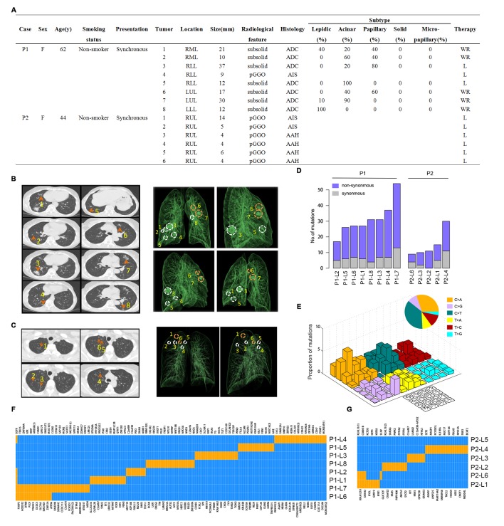
Radiological features and whole-exome sequencing of the two patients. (A) Clinicopathological characteristics. RML, right middle lobe; RLL, right lower lobe; LUL, left upper lobe; LLL, left lower lobe; RUL, right upper lobe; pGGO, pure ground-glass opacities; ADC, adenocarcinoma; AIS, adenocarcinoma in situ; AAH, atypical adenomatous hyperplasia; WR, wedge resection; L, lobectomy; (B) Left: chest CT scans obtained with 1 mm thick sections of patient 1 (P1) with eight scattered GGOs (orange arrows) in the bilateral lung. Right: reconstructed lung 3D images of P1. Circles denote the spatial locations of the lesions and orange circles indicate metastatic lesions. (C) Left: CT scans obtained with 1 mm thick sections of patient 2 (P2) with six scattered GGOs (orange arrows) in her right upper lobe. All six lesions are pure GGOs of very small size. Right: reconstructed 3D images of P2. Circles denote the spatial locations of the lesions and orange circles indicate metastatic lesions. (D) Number of somatic mutations identified in each lesion of the two patients. Lesion name is in form of ‘Patient ID + lesion number’. For example, P1-L1 stands for the number 1 lesion of patient 1. (E) Mutational signature of P1 based on all somatic mutations detected in this patient. (F) Regional distribution of non-synonymous somatic mutations among the eight lesions of P1. Each column represents a single mutation site. Blue represents wild type while orange represents mutation in a certain site of a certain gene. (G) Regional distribution of somatic mutations among the six lesions of P2. Each column represents a single mutation site. Blue represents wild type while orange represents mutation in a certain site of a certain gene.
Comment in
-
Multifocal adenocarcinoma: perspectives, assumptions and elephants.J Thorac Dis. 2018 Mar;10(3):1193-1197. doi: 10.21037/jtd.2018.01.173. J Thorac Dis. 2018. PMID: 29708150 Free PMC article. No abstract available.
-
Multifocal ground-glass opacities: multifocal origin versus intrapulmonary metastasis.J Thorac Dis. 2018 Mar;10(3):1253-1255. doi: 10.21037/jtd.2018.03.25. J Thorac Dis. 2018. PMID: 29708176 Free PMC article. No abstract available.
References
-
- Detterbeck FC, Marom EM, Arenberg DA, et al. The IASLC lung cancer staging project: background data and proposals for the application of TNM staging rules to lung cancer presenting as multiple nodules with ground glass or lepidic features or a pneumonic type of involvement in the forthcoming eighth edition of the TNM classification. J Thorac Oncol 2016;11:666–80. 10.1016/j.jtho.2015.12.113 - DOI - PubMed
-
- Kadota K, Nitadori J, Sima CS, et al. Tumor spread through air spaces is an important pattern of invasion and impacts the frequency and location of recurrences after limited resection for small stage I lung adenocarcinomas. J Thorac Oncol 2015;10:806–14. 10.1097/JTO.0000000000000486 - DOI - PMC - PubMed
Publication types
MeSH terms
LinkOut - more resources
Full Text Sources
Other Literature Sources
Medical