Case Reports
. 2017 Sep 19;4(3):46.
doi: 10.3390/vetsci4030046.

Infection by Microsporum canis in Paediatric Patients: A Veterinary Perspective

Affiliations
Case Reports

Infection by Microsporum canis in Paediatric Patients: A Veterinary Perspective

Mario Pasquetti et al. Vet Sci. .

Abstract

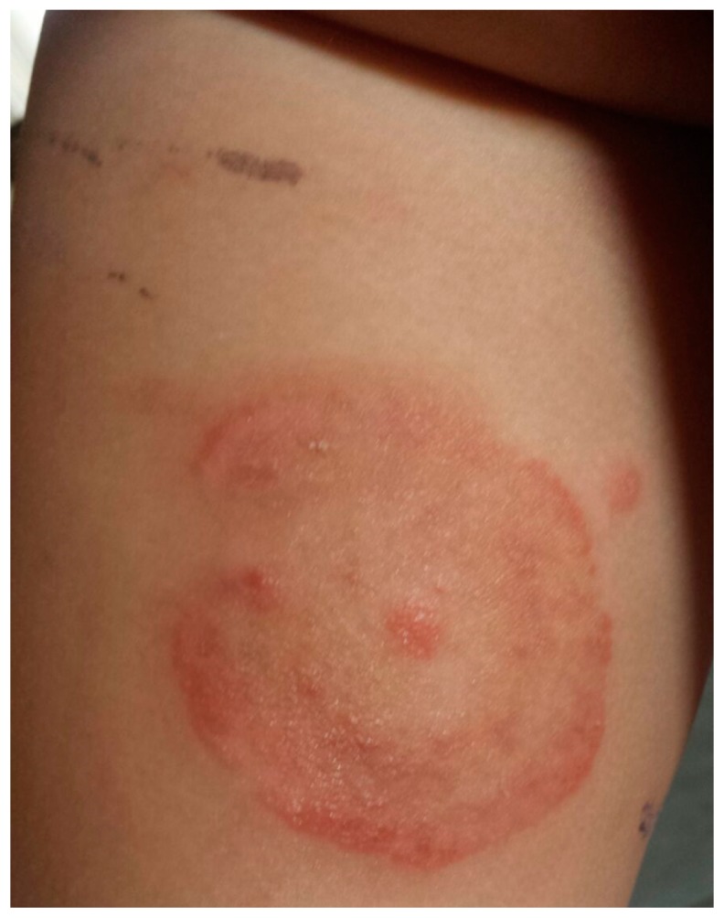
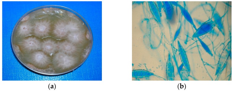
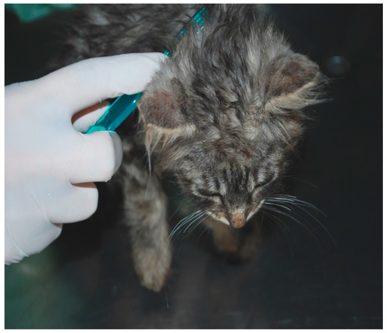
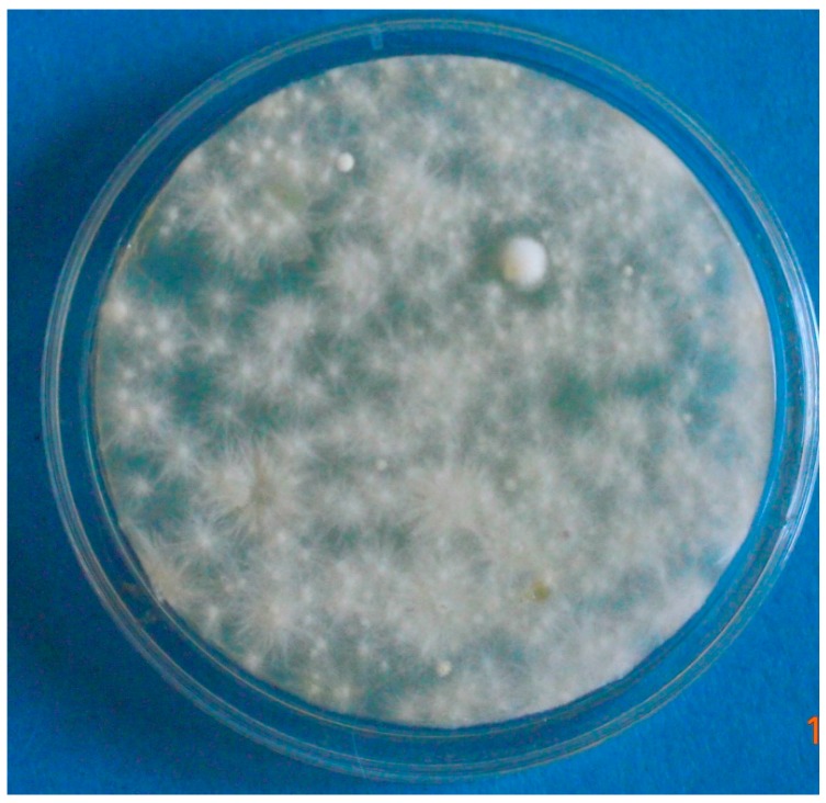
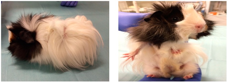
Microsporum canis is a dermatophyte fungus of which cats and dogs are recognized as the natural hosts. M. canis is also easily transmitted to humans, causing lesions to the glabrous skin (tinea corporis) and to the head (tinea capitis). The present study describes some cases of infection with M. canis in children from a veterinary perspective, highlighting some important features of this clinical entity (e.g., the necessity to identify the animal source of infection with appropriate diagnostic tests; the fact that infected cats may present with no or atypical dermatological signs; and the importance of the environment as a fungal reserve).

Keywords: Microsporum canis; cat; dermatophytes; paediatric; ringworm.


Conflict of interest statement

The authors declare that there are no conflicts of interest.

Figures

Figure 1
Figure 1
Tinea corporis due to M. canis in a child (case # 3).
Figure 2
Figure 2
(a) Colonies of M. canis in culture; (b) Microscopic aspect, macro- and micro-conidia.
Figure 3
Figure 3
A kitten infected by M. canis (source of infection in case # 1) with no evidence of clinical lesions, sampled by the tooth-brush technique.
Figure 4
Figure 4
Environmental sample (contact plate from sofa, case # 1). Widespread growth of colonies of M. canis.
Figure 5
Figure 5
A guinea pig presented as the probable cause of infection by M. canis in a child (case # 3).

References

    1. Weitzman I., Summerbell R.C. The dermatophytes. Clin. Microbiol. Rev. 1995;8:240–259. doi: 10.1016/S0733-8635(05)70320-X. - DOI - PMC - PubMed
    1. Chermette R., Ferreiro L., Guillot J. Dermatophytoses in Animals. Mycopathologia. 2008;166:385–405. doi: 10.1007/s11046-008-9102-7. - DOI - PubMed
    1. Gallo M.G., Tizzani P., Peano A., Rambozzi L., Meneguz P.G. Eastern cottontail (Sylvilagus floridanus) as carrier of dermatophyte fungi. Mycopathologia. 2005;160:163–166. doi: 10.1007/s11046-005-6619-x. - DOI - PubMed
    1. Gallo M.G., Lanfranchi P., Poglayen G., Calderola S., Menzano A., Ferroglio E., Peano A. Seasonal 4-year investigation into the role of the alpine marmot (Marmota marmota) as a carrier of zoophilic dermatophytes. Med. Mycol. 2005;43:373–379. doi: 10.1080/13693780400008282. - DOI - PubMed
    1. Pal M., Dave P. Tinea faciei in a goat handler due to Microsporum canis. Rev. Iberoam. Micol. 2005;22:181–182. doi: 10.1016/S1130-1406(05)70039-5. - DOI - PubMed

Publication types