

Establishment and Characterization of New Canine and Feline Osteosarcoma Primary Cell Lines
- PMID: 29056719
- PMCID: PMC5644629
- DOI: 10.3390/vetsci3020009
Establishment and Characterization of New Canine and Feline Osteosarcoma Primary Cell Lines
Abstract
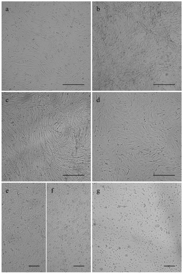
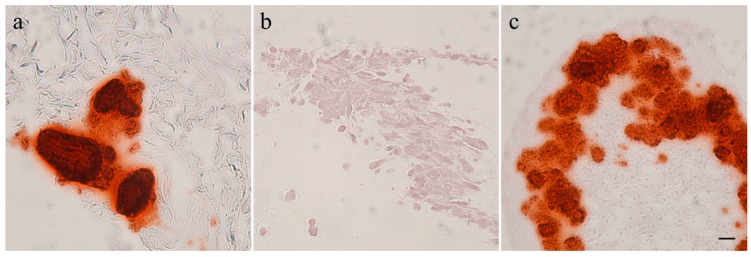
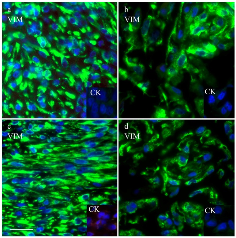
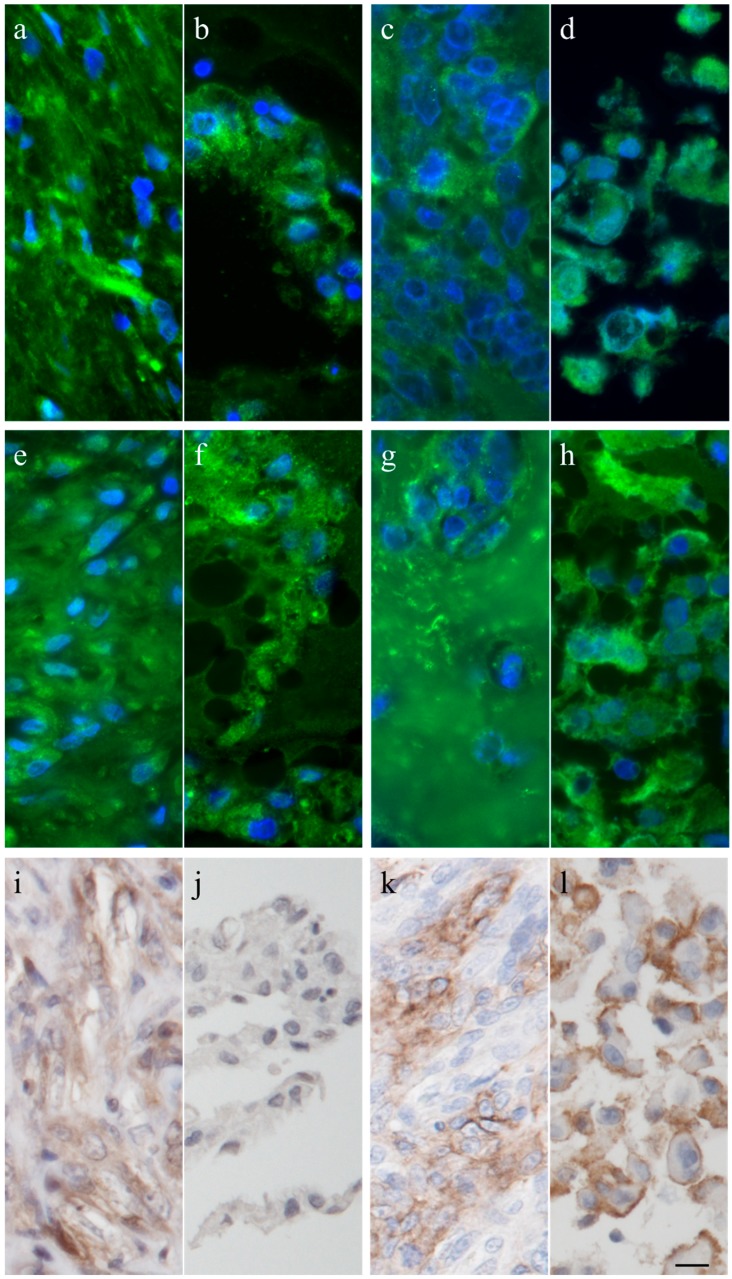
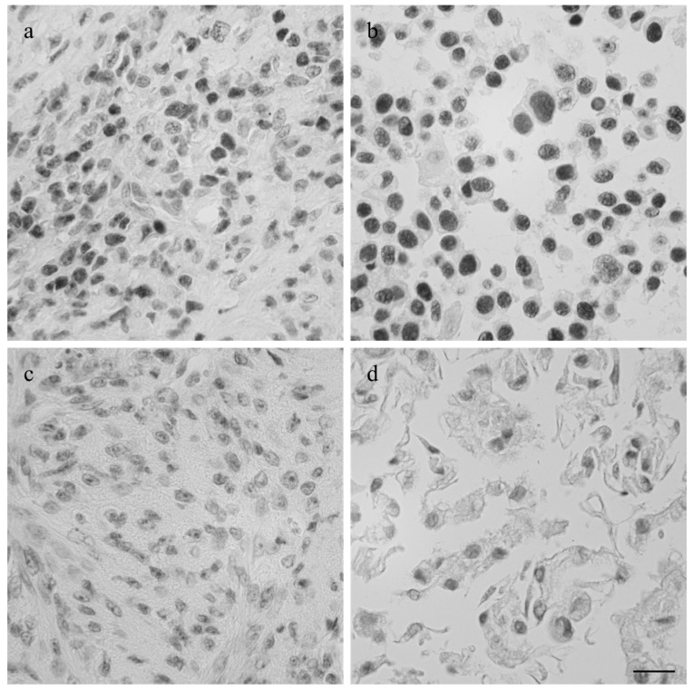
Osteosarcomas are the most abundant form of bone malignancies in multiple species. Canine osteosarcomas are considered a valuable model for human osteosarcomas because of their similar features. Feline osteosarcomas, on the other hand, are rarely studied but have interesting characteristics, such as a better survival prognosis than dogs or humans, and less likelihood of metastasis. To enable experimental approaches to study these differences we have established five new canine osteosarcoma cell lines out of three tumors, COS_1186h, COS_1186w, COS_1189, and COS_1220, one osteosarcoma-derived lung metastasis, COS_1033, and two new feline osteosarcoma cell lines, FOS_1077 and FOS_1140. Their osteogenic and neoplastic origin, as well as their potential to produce calcified structures, was determined by the markers osteocalcin, osteonectin, tissue unspecific alkaline phosphatase, p53, cytokeratin, vimentin, and alizarin red. The newly developed cell lines retained most of their markers in vitro but only spontaneously formed spheroids produced by COS_1189 showed calcification in vitro.
Keywords: cat; cell culture; dog; osteosarcoma.
Conflict of interest statement
The authors declare no conflict of interest.
Figures
References
-
- World Cancer Research Fund International. [(accessed on 31 May 2016)]. Available online: https://www.wcrf.org/int/cancer-facts-figures/data-cancer-frequency-country.
-
- Dorn C.R., Taylor D.O., Schneider R., Hibbard H.H., Klauber M.R. Survey of animal neoplasms in Alameda and Contra Costa Counties, California. II. Cancer morbidity in dogs and cats from Alameda County. J. Natl. Cancer Inst. 1968;40:307–318. - PubMed
-
- Boerma M., Burton G.R., Wang J., Fink L.M., McGehee R.E., Hauer-Jensen M. Comparative expression profiling in primary and immortalized endothelial cells: Changes in gene expression in response to hydroxy methylglutaryl-coenzyme A reductase inhibition. Blood Coagul. Fibrinolysis. 2006;17:173–180. doi: 10.1097/01.mbc.0000220237.99843.a1. - DOI - PubMed
Grants and funding
LinkOut - more resources
Full Text Sources
Other Literature Sources
Research Materials
Miscellaneous
