

Engineered Peptide Repairs Defective Adhesive-Dentin Interface
- PMID: 29056869
- PMCID: PMC5650097
- DOI: 10.1002/mame.201600487
Engineered Peptide Repairs Defective Adhesive-Dentin Interface
Abstract
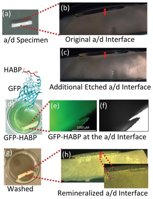
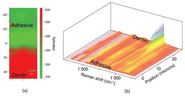
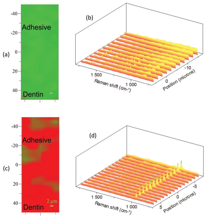
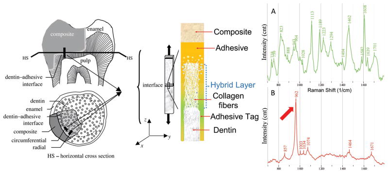
Failure of dental composite restorations is primarily due to recurrent decay at the tooth-composite interface. At this interface, the adhesive and its bond with dentin is the barrier between the restored tooth and the oral environment. In vivo degradation of the bond formed at the adhesive/dentin (a/d) interface follows a cascade of events leading to weakening of the composite restoration. Here, a peptide-based approach is developed to mineralize deficient dentin matrices at the a/d interface. Peptides that have an inherent capacity to self-assemble on dentin and to induce calcium-phosphate remineralization are anchored at the interface. Distribution of adhesive, collagen, and mineral is analyzed using micro-Raman spectroscopy and fluorescence microscopy. The analysis demonstrates remineralization of the deficient dentin matrices achieved throughout the interface with homogeneous distribution of mineral. The peptide-based remineralization demonstrated here can be an enabling technology to design integrated biomaterial-tissue interfaces.
Keywords: adhesive/dentin interface; composite restorative materials; material–tissue interface; mineralization; peptides.
Figures
References
-
- DeRouen TA, Martin MD, Leroux BG, Townes BD, Woods JS, Leitao J, Castro-Caldas A, Luis H, Bernardo M, Rosenbaum G, Martins IP. JAMA, J Am Med Assoc. 2006;295:1784. - PubMed
-
- Simecek JW, Diefenderfer KE, Cohen ME. J Am Dent Assoc. 2009;140:200. - PubMed
-
- van de Sande FH, Opdam NJ, Rodolpho PAD, Correa MB, Demarco FF, Cenci MS. J Dent Res. 2013;92:S78. - PubMed
-
- Bernardo M, Luis H, Martin MD, Leroux BG, Rue T, Leitao J, DeRouen TA. J Am Dent Assoc. 2007;138:775. - PubMed
Grants and funding
LinkOut - more resources
Full Text Sources
Other Literature Sources