. 2017 Jun;5(2):145-152.
doi: 10.1007/s40139-017-0132-z. Epub 2017 Apr 22.

Mechanisms of Fibroblast Activation in the Remodeling Myocardium

Affiliations

Mechanisms of Fibroblast Activation in the Remodeling Myocardium

Arti V Shinde et al. Curr Pathobiol Rep. 2017 Jun.

Abstract

Purpose of review: Activated fibroblasts are critically implicated in repair and remodeling of the injured heart. This manuscript discusses recent progress in the cell biology of fibroblasts in the infarcted and remodeling myocardium, highlighting advances in understanding the origin, function and mechanisms of activation of these cells.

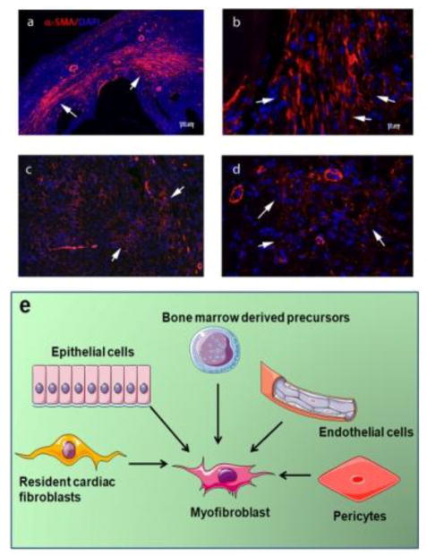
Recent findings: Following myocardial injury, fibroblasts undergo activation and myofibroblast transdifferentiation. Recently published studies have suggested that most activated myofibroblasts in the infarcted and pressure-overloaded hearts are derived from resident fibroblast populations. In the healing infarct, fibroblasts undergo dynamic phenotypic alterations in response to changes in the cytokine milieu and in the composition of the extracellular matrix. Fibroblasts do not simply serve as matrix-producing cells, but may also regulate inflammation, modulate cardiomyocyte survival and function, mediate angiogenesis, and contribute to phagocytosis of dead cells.

Summary: In the injured myocardium, fibroblasts are derived predominantly from resident populations and serve a wide range of functions.

Keywords: cardiac remodeling; cytokine; fibroblast; myocardial infarction; myofibroblast.


Figures

Figure 1
Figure 1. Myofibroblasts in the infarcted and remodeling myocardium
αSMA immunofluorescence identifies abundant myofibroblasts (arrows) in infarcted mouse hearts (after 7 days of coronary occlusion) (a, b) and in pressure overloaded hearts after 7 days of transverse aortic constriction (c, d). (e) Myofibroblasts in injured hearts may originate from a variety of sources, including epicardial epithelial cells, endothelium (through EndMT), vascular pericytes, bone marrow derived precursors, and resident cardiac fibroblasts. Recently published studies in mouse models using lineage tracing approaches suggest that resident cardiac fibroblast populations may be the most important source of activated myofibroblasts in infarcted and pressure-overloaded hearts.

References

    1. Christiansen MN, Kober L, Weeke P, Vasan RS, Jeppesen JL, Smith JG, et al. Age-specific Trends in Incidence, Mortality and Comorbidities of Heart Failure in Denmark 1995–2012. Circulation. 2017 doi: 10.1161/CIRCULATIONAHA.116.025941. - DOI - PubMed
    1. Chen J, Hsieh AF, Dharmarajan K, Masoudi FA, Krumholz HM. National trends in heart failure hospitalization after acute myocardial infarction for Medicare beneficiaries: 1998–2010. Circulation. 2013;128(24):2577–84. doi: 10.1161/CIRCULATIONAHA.113.003668. - DOI - PMC - PubMed
    1. Kong P, Christia P, Frangogiannis NG. The pathogenesis of cardiac fibrosis. Cell Mol Life Sci. 2014;71(4):549–74. - PMC - PubMed
    1. Berk BC, Fujiwara K, Lehoux S. ECM remodeling in hypertensive heart disease. J Clin Invest. 2007;117(3):568–75. - PMC - PubMed
    1. Shinde AV, Frangogiannis NG. Fibroblasts in myocardial infarction: A role in inflammation and repair. J Mol Cell Cardiol. 2014;70C:74–82. - PMC - PubMed

LinkOut - more resources