Review
. 2018 Jan 2;47(1):28-52.
doi: 10.1039/c7cs00557a.

Fluorogenic reaction-based prodrug conjugates as targeted cancer theranostics

Affiliations
Review

Fluorogenic reaction-based prodrug conjugates as targeted cancer theranostics

Min Hee Lee et al. Chem Soc Rev. .

Abstract

Theranostic systems are receiving ever-increasing attention due to their potential therapeutic utility, imaging enhancement capability, and promise for advancing the field of personalized medicine, particularly as it relates to the diagnosis, staging, and treatment of cancer. In this Tutorial Review, we provide an introduction to the concepts of theranostic drug delivery effected via use of conjugates that are able to target cancer cells selectively, provide cytotoxic chemotherapeutics, and produce readily monitored imaging signals in vitro and in vivo. The underlying design concepts, requiring the synthesis of conjugates composed of imaging reporters, masked chemotherapeutic drugs, cleavable linkers, and cancer targeting ligands, are discussed. Particular emphasis is placed on highlighting the potential benefits of fluorogenic reaction-based targeted systems that are activated for both imaging and therapy by cellular entities, e.g., thiols, reactive oxygen species and enzymes, which are present at relatively elevated levels in tumour environments, physiological characteristics of cancer, e.g., hypoxia and acidic pH. Also discussed are systems activated by an external stimulus, such as light. The work summarized in this Tutorial Review will help define the role fluorogenic reaction-based, cancer-targeting theranostics may have in advancing drug discovery efforts, as well as improving our understanding of cellular uptake and drug release mechanisms.


Conflict of interest statement

The authors declare no competing financial interest.

Figures

Fig. 1
Fig. 1
Design principle for achieving fluorogenic reaction-based prodrug conjugates that are able to target cancer cells selectively, provide cytotoxic chemotherapeutics, and produce readily-monitored imaging signals in vitro and in vivo.
Fig. 2
Fig. 2
GSH-induced disulfide bond cleavage of theranostic agent 1. The Ex1/Em1 and Ex2/Em2 represent, respectively, the excitation and emission wavelengths before and after therapeutic activation.
Fig. 3
Fig. 3
(A) Thiol-activatable theranostic agent FL-2 and controls L-2 and FL-3. (B) Proposed drug release mechanism and fluorescence turn on features expected for FL-2 upon reaction with cellular thiols.
Fig. 4
Fig. 4
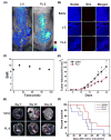
Bioimaging and therapeutic effects of theranostic FL-2 and controls L-2 and FL-3 in (A–D) xenograft tumour nude mice and (E & F) metastatic liver cancer mice. (A–F) are reproduced with permission from ref. 12. Copyright 2016 American Chemical Society.
Fig. 5
Fig. 5
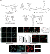
Thiol-activatable theranostic agents 4–9.
Fig. 6
Fig. 6
Thiol-activatable NIR dye-based theranostic agents 10–15. Also shown is a NIR dye used for in vivo imaging, ICG.
Fig. 7
Fig. 7
(A) Thiol-activatable NIR dye-based theranostic agents 16 and 17, and its disulfide cleavage reaction. (B–D) In vivo and (E–J) ex vivo biodistribution images of mice treated with agents 16, 17, and CyA-K. (B–J) are reproduced from ref. 24 with permission from the Royal Society of Chemistry.
Fig. 8
Fig. 8
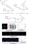
(A) Thiol-activatable AIE-based theranostic agents 18 and 19. (B) Conjugate 20 and its activation by GSH and cathepsin B, respectively.
Fig. 9
Fig. 9
(A) H2O2-triggered theranostic systems (21–23). (B–D) Bio-imaging and therapeutic effects of theranostics 21, 22, and 23 respectively. (Bi-iii) in vivo MRI images of normal mice and saline, 21 treated lung metastasis mice (axial plane views) at day 10 post-inoculation, respectively, and (Biv-vi) images of lungs isolated from the mice. (Ci) mitochondrial ultrastructure after treatment with 22 in LPS pre-treated cells. (Di) whole body image of U-87 MG tumour bearing mice after 23 injection (1 min) and (Dii) ex vivo images of dissected organs 5 min post injection. (B) and (C) are reproduced with permission from ref. 27 and 28. Copyright 2014 American Chemical Society. (D) was adapted from ref. 29. Copyright 2015 Wiley-VCH.
Fig. 10
Fig. 10
(A) Acid-activated theranostic systems 24–27. (B, C, D and E) Bio-imaging and therapeutic effect of theranostic 24, 25, 26 and 27 respectively. (Ci-iv) confocal microscopy images of U87 cells upon treatment with 25 (post 42 h) and cell viability at different concentration of 25, free Dox and control. Cell nuclei stained with Hoechst 33342 (blue), Dox (red fluorescence), FAM (blue fluorescence), and merged image. (Di) Normalized cell viability of 26 to nonactivated (media), LPS-, and IL-4-treated macrophages. (Dii & Diii) Live fluorescence confocal microscopic in vivo imaging of macrophages in 26-treated zebrafish without (Dii) and with (Diii) LPS treatment. The arrow heads indicate the Dox activation (green) and surrounding apoptotic macrophages (red). (Ei-iii) confocal images of CT26 cells pre-treated with Hoechst (blue), Lysotracker (green) and 27 (red). (Eiv) T1-weighted MR images of A549 and CT26 cells co-incubated with 27 at various concentrations at 200 MHz. (B–E) are reproduced with permission from references 31, 32, 33, and 34, respectively. Copyright 2014 Royal Society of Chemistry (for B), Copyright 2015 Wiley-VCH (for C), 2017 American Chemical Society (for D), and Copyright 2016 Royal Society of Chemistry (for E).
Fig. 11
Fig. 11
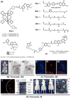
(A) Hypoxia-activated theranostic agent 28 and its cleavage reaction. (B) Chemical structure of the reference compounds 28a and 28b. (C–F) Bio-imaging and therapeutic effects of theranostic 28. (C–F) are reproduced with permission from ref. 35. Copyright 2016 Elsevier.
Fig. 12
Fig. 12
Design strategy underlying theranostic 29, an agent designed to release the cytotoxic species GMC via controlled UV-irradiation under hypoxic conditions. An active coumarin fluorophore is also produced.
Fig. 13
Fig. 13
(A) Hypoxia-activated theranostic agent 30 and its mode of cleavage. (B–G) Bio-imaging and therapeutic effects of theranostic 30. (B–G) are reproduced with permission from ref. 37. Copyright 2017 Elsevier.
Fig. 14
Fig. 14
(A) Platinum-based theranostic agents (31–33). (B–E) Bio-imaging and therapeutic response of theranostic agents 31, 32, and 33 respectively. (Bi-iv) confocal images of MDA-MB-231 cells pre-treated with 31 at 1 h, 2 h, 4 h and 6 h, respectively, and (Bv) cell viability upon treatment with 31 and other controls. (Di-iv) real time confocal images for apoptosis progress in U87-MG cells stained with 32 and MCF-67 cells (Dv) and 293T cells (Dvi). (Ei-iii & iv) confocal images of MDA-MB-231 cells and U87-MG cells recorded after incubation with 33 at different time intervals. (Ev) viability of U87-MG and MDA-MB-231 cells upon incubation with 33 in the dark and with light illumination (0.25 W cm−2 for 1 min). (B–E) are reproduced with permission from refs. 38, 39, and 40, respectively. Copyright 2014 Royal Society of Chemistry (for B), Copyright 2014 American Chemical Society (for C and D), and Copyright 2015 Royal Society of Chemistry (for E).
Fig. 15
Fig. 15
(A) Design of theranostic agent 34 and its activation. (B) Enzyme-activatable theranostic agents (34a-34c). (C) Enzymatic activation of theranostic agent 34d.
Fig. 16
Fig. 16
Proposed enzymatic activation of theranostic agent 35.
Fig. 17
Fig. 17
(A) Enzymatic activation of theranostic agent 36. (B) Fluorescence response seen when 36 was allowed to react with DT-diaphorase (NQO1). (C–G) Bioimaging and therapeutic effects of 36. (B–G) are reproduced with permission from ref. 43. Copyright 2016 American Chemical Society.
Fig. 18
Fig. 18
Enzymatic activation of theranostic agent 37.
Fig. 19
Fig. 19
(A) Light-activated prodrugs 38–40. (B–C) Bio-imaging and therapeutic effects of 38d, 39b, and 40. (Bi) Fluorescence image of SC colon cancer bearing Balb/c mice after 38d treatment (7 h post injection) and (Bii & iii) photographic images of mice treated with 38d and 38e and illumination (690 nm, 100 mW cm−2, 30 min) at day 15 post-illumination. (Ci-iii) fluorescence images of HeLa cells recorded after UV irradiation (30 min) and pre-treated with 39b showing blue fluorescence (activated 39b), red (propidium iodide stained nuclei), and merged images respectively. (Di) fluorescence image of A549 cells pre-treated with 40 before and after illumination at 405 nm (1 h). (Dii) Enhanced expression of various apoptosis gene/markers upon treatment with 40 and illumination under different conditions in A549 cells. (Diii & iv) In vivo images of A549 bearing mice injected with 40 and control with/without illumination and ex vivo images of tumour and organs after treatment with 40 and photoillumination. (B–D) are reproduced with permission from ref. 48, 49, and 50, respectively. Copyright 2014 American Chemical Society (for B), Copyright 2015 Royal Society of Chemistry (for C), and Copyright 2016 Nature Publishing Group (for D).

References

    1. Kumar R, Shin WS, Sunwoo K, Kim WY, Koo S, Bhuniya S, Kim JS. Chem. Soc. Rev. 2015;44:6670. - PubMed
    1. Lim EK, Kim T, Paik S, Haam S, Huh Y-M, Lee K. Chem. Rev. 2015;115:327. - PubMed
    1. Weinstain R, Segal E, Satchi-Fainaro R, Shabat D. Chem. Commun. 2010;46:553. - PubMed
    1. Lee MH, Sessler JL, Kim JS. Acc. Chem. Res. 2015;48:2935. - PubMed
    1. Gnaim S, Shabat D. Acc. Chem. Res. 2014;47:2970. - PubMed