. 2017 Oct 20;7(1):13652.
doi: 10.1038/s41598-017-13947-3.

Alcohol consumption impairs the ependymal cilia motility in the brain ventricles

Affiliations

Alcohol consumption impairs the ependymal cilia motility in the brain ventricles

Alzahra J Al Omran et al. Sci Rep. .

Abstract

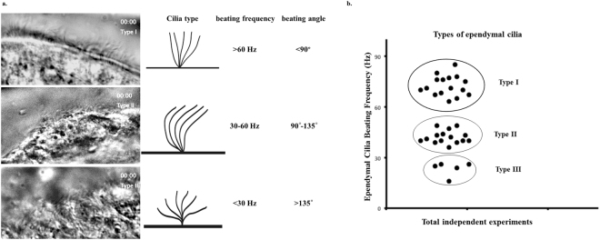
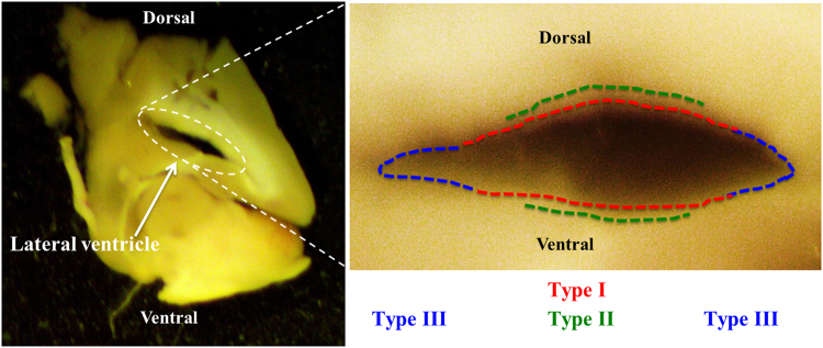
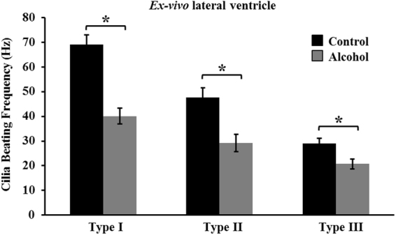
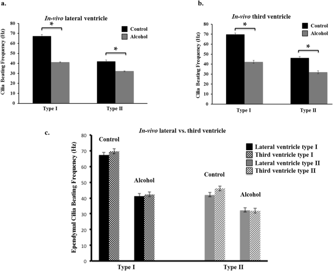
Ependymal cilia protrude into the central canal of the brain ventricles and spinal cord to circulate the cerebral spinal fluid (CSF). Ependymal cilia dysfunction can hinder the movement of CSF leading to an abnormal accumulation of CSF within the brain known as hydrocephalus. Although the etiology of hydrocephalus was studied before, the effects of ethanol ingestion on ependymal cilia function have not been investigated in vivo. Here, we report three distinct types of ependymal cilia, type-I, type-II and type-III classified based upon their beating frequency, their beating angle, and their distinct localization within the mouse brain-lateral ventricle. Our studies show for the first time that oral gavage of ethanol decreased the beating frequency of all three types of ependymal cilia in both the third and the lateral rat brain ventricles in vivo. Furthermore, we show for the first time that hydin, a hydrocephalus-inducing gene product whose mutation impairs ciliary motility, and polycystin-2, whose ablation is associated with hydrocephalus are colocalized to the ependymal cilia. Thus, our studies reinforce the presence of three types of ependymal cilia in the brain ventricles and demonstrate the involvement of ethanol as a risk factor for the impairment of ependymal cilia motility in the brain.


Conflict of interest statement

The authors declare that they have no competing interests.

Figures

Figure 1
Figure 1
Ependymal cilia of the brain lateral ventricle are classified into three types based on their beating frequency and angle. (a) Shown here are representative DIC images of the ependymal cells of the mouse brain lateral ventricle with the three types of motile cilia. The images of the ependymal cilia are taken from a time-lapse movie during a live imaging experiment. (b) Classification of ependymal cilia is based upon their beating frequency and angle. Each dot represents an independent experiment. Type I cilia are the fastest and has a beating frequency >60 Hz with a beating angle less than 90°. Type II beating frequency is between 30–60 Hz with a beating angle between 90°–135°. Type III cilia have the slowest beating frequency <30 Hz and a beating angle >135°.
Figure 2
Figure 2
Each type of ependymal cilia has specific localization within the brain lateral ventricle. This figure shows a sagittal section view of the lateral ventricle (left). Each type of the ependymal cilia (shown on the enlarged area) is localized within specific area in the lateral ventricle based on the beating frequencies and angle of movement.
Figure 3
Figure 3
Ethanol decreased cilia beating ex vivo. Treatment of ex vivo brain slices with 0.25% ethanol for five minutes shows a significant reduction in the beating frequency of all three types of ependymal cilia. Up to 22 independent preparations were used and the presence of an asterisk (*) denotes significant difference at p < 0.05.
Figure 4
Figure 4
Alcohol drinking altered the dynamics of ependymal cilia in the brain. Two-month old Wistar rats were given either water or 95% ethanol (alcohol) at a 6 g per kg of body weight for seven days. After acute oral treatment with alcohol, the rat’s brain was dissected to examine the dynamics of ependymal cilia in both the lateral and third ventricles. Alcohol drinking caused a significant decrease in the beating frequency of the ependymal cilia of both, (a) the lateral and (b) third ventricles compared to the control group. (c) Further comparison within the control groups and the alcohol-drinking groups between the dynamics of the ependymal cilia in the lateral ventricle vs. the third ventricle demonstrated no significant difference between the ciliary beating frequencies.

Comment in

References

    1. Banizs B, et al. Dysfunctional cilia lead to altered ependyma and choroid plexus function, and result in the formation of hydrocephalus. Development (Cambridge, England) 2005;132:5329–5339. doi: 10.1242/dev.02153. - DOI - PubMed
    1. Jimenez AJ, Dominguez-Pinos MD, Guerra MM, Fernandez-Llebrez P, Perez-Figares JM. Structure and function of the ependymal barrier and diseases associated with ependyma disruption. Tissue barriers. 2014;2:e28426. doi: 10.4161/tisb.28426. - DOI - PMC - PubMed
    1. Clarren SK, Alvord EC, Jr., Sumi SM, Streissguth AP, Smith DW. Brain malformations related to prenatal exposure to ethanol. J. Pediatr. 1978;92:64–67. doi: 10.1016/S0022-3476(78)80072-9. - DOI - PubMed
    1. Kahle KT, Kulkarni AV, Limbrick DD, Warf BC. Hydrocephalus in children. Lancet. 2016;387:788–799. doi: 10.1016/S0140-6736(15)60694-8. - DOI - PubMed
    1. Al-Dosari MS, et al. Mutation in MPDZ causes severe congenital hydrocephalus. J. Med. Genet. 2013;50:54–58. doi: 10.1136/jmedgenet-2012-101294. - DOI - PubMed

Publication types

MeSH terms