. 2017 Oct 20;7(1):13663.
doi: 10.1038/s41598-017-13961-5.

Energetic mitochondrial failing in vitiligo and possible rescue by cardiolipin

Affiliations

Energetic mitochondrial failing in vitiligo and possible rescue by cardiolipin

Maria Lucia Dell'Anna et al. Sci Rep. .

Abstract

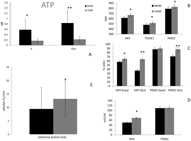
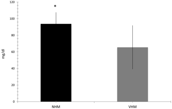
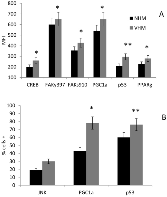
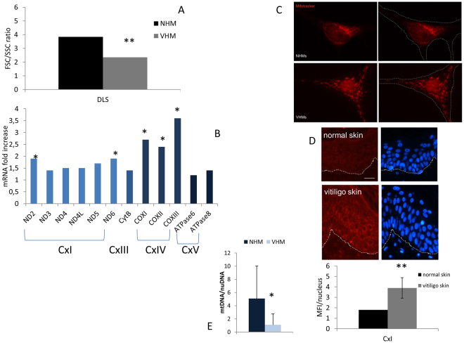
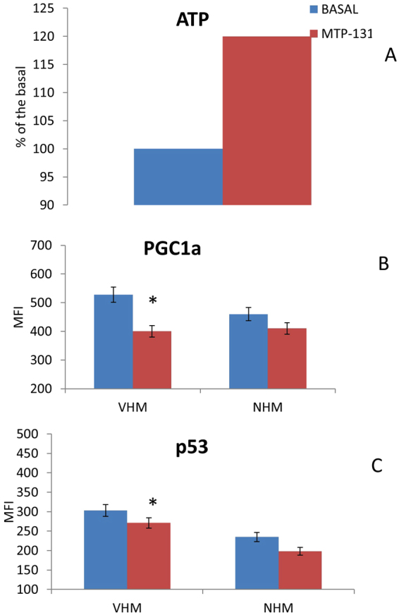
Vitiligo is characterized by death or functional defects of epidermal melanocytes through still controversial pathogenic process. Previously, we showed that mitochondria-driven pre-senescent phenotype diminishes the capability of vitiligo melanocytes to cope with stressful stimuli. In the current study, we investigated markers of mitochondrial energy metabolism including the PGC1a axis, and then we determined the index of mitochondrial impairment using a cytomic approach. We found in cultured epidermal vitiligo melanocytes, compared to healthy ones, low ATP, increased proton leakage, and altered expression of several glycolytic enzymes (hexokinase II, pyruvic dehydrogenase kinase 1 and pyruvic kinase M2), We suggest that the low ATP production may be sufficient in steady-state conditions but it is unable to cover further needs. We also found in vitiligo melanocyrtes hyper-activation of the PGC1α axis, finalized to counteract the energy defect. Cytomic analysis, supported by MitoTracker Red pattern and ex-vivo immunohistochemistry, suggested an increased mitochondrial mass, possibly useful to ensure the essential ATP level. Finally, pharmacological cardiolipin stabilization reverted the energetic impairment, confirming the initial mitochondrial role. In conclusion, we report new insight in the pathogenetic mechanism of viitligo and indicate that the mitochondrial failure rescue by cardiolipin manipulation may be a new intriguing target in treatment development.


Conflict of interest statement

The Authors declared the occurrence of Conflct of Interest. DA Brown has received research grants and has served as a consultant for Stealth BioTherapeutics. The research was supported by unrestricted grants from Stealth Biotherapeutics. DA Brown has received research grants and has served as a consultant for Stealth BioTherapeutics.

Figures

Figure 1
Figure 1
ATP production in NHM and VHM in basal and forced mitochondrial activity. (A) Quantification has been performed by fluorimetric kit. It was carried out in basal condition (standard culture medium) and after 12 days treatment with 20 mM L-glutamine. Data are mean ± SD of all the performed experiments. *p = 0.008; **p = 0.009. (B) Flow cytometric analysis of MFI for HKII, PKM2, and PDHK1. (C) The percentage of positive cells for HKII and PKM2 was also evaluated and an increased expression was observed in VHM in basal condition; NHM down-regulate its expression after L-glutamine supplementation whereas VHM aren’t affected by the presence of the alternative substrate, underlying the difference between the two populations. (D) The activity of HKII was also forced in a compensatory strategy. Data are mean ± SD of all the performed experiments. *p = 0.05; **p = 0.01. (E) A relevant proton leak was detected in VHM, underlying the inability of ETC to correctly finalize the specific activity. Consequently, the mitochondrial performance, represented by BHI, was negatively affected. BHI is calculated as (ATP × Reserve Capacity)/(proton leak × non mitochondrial respiration). Data are mean ± SD of all the performed experiments. *p = 0.01.
Figure 2
Figure 2
Glucose uptake. Spectrophotometric assay defined increased glucose uptake by the defined medium according to the increased expression and activity of HKII. Data are mean ± SD of all the performed experiments. *p = 0.05.
Figure 3
Figure 3
How the cross-talk mitochondrion-nucleus may be affected. Flow cytometric analysis of some key factors of the intracellular network confirmed the increased phosphorylation of CREB and revealed a parallel higher phosphorylation of FAK which, together with the increased expression of PGC1a p53 and PPARg, testified for the increased mitogenesis in an attempt to improve mitocohondrial activitiy. Both MFI and percentage of positive cells were increased in VHM population. Results are referred to analyses carried out in basal condition. Data are mean ± SD of all the performed experiments. *p = 0.05; **p = 0.01.
Figure 4
Figure 4
Multiparametric analysis of mitochondrial mass. (A) Cytomic approach, based on the DLS parameter, demonstrated increased mitochondrial mass. It was further explored by alternative methods: (B) mRNA for some mitochondrial proteins was increased; (C) analysis of Mitotracker signal by fluorescence microscope underlined an enlarged structure and a specific organization of mitochondria in VHM; (D) signal for CxI expression obtained by immunohistochemistry staining was increased. Finally, (E) mtDNA quantitation, performed using nuclear DNA as internal reference, confirmed the increased volume of mitochondria. Data are mean ± SD of all the performed experiments. *p = 0.02; **p = 0.05.
Figure 5
Figure 5
Cardiolipin stabilization may improve mitochondrial performance. The in vitro treatment of cells with 2μM MTP-131 for 7 days rescued the ability of VHM to (A) produce ATP. Consequently, the expression of (B) PGC1a and (C) p53 was reduced. Data are mean ± SD of all the performed experiments. *p = 0.05.

References

    1. Schallreuter KU, et al. Vitiligo pathogenesis: autoimmune disease, genetic defect, excessive reactive oxygen species, calcium imbalance, or what else? Exp, Dermatol. 2008;17:139–160. doi: 10.1111/j.1600-0625.2007.00666.x. - DOI - PubMed
    1. Picardo, M. et al. Vitiligo. Nat. Rev. Dis. Primers 1, 15011, 10.1038/nrdp.2015.11 (2015). - PubMed
    1. Strassner JP, Harris JE. Understanding mechanisms of autoimmunity through translational research in vitiligo. Curr Opin Immunol. 2016;43:81–88. doi: 10.1016/j.coi.2016.09.008. - DOI - PMC - PubMed
    1. Jin Y, et al. Genome-wide association studies of autoimmune vitiligo identify 23 new risk loci and highlight pathways and regulatory variants. Nat, Genet. 2016;48:1418–1424. doi: 10.1038/ng.3680. - DOI - PMC - PubMed
    1. Picardo, M, Taieb, A (Eds). Vitiligo, Springer Ed (2010).

Publication types