. 2018:1684:223-235.
doi: 10.1007/978-1-4939-7362-0_17.

Stopped-Flow Fluorometric Ion Flux Assay for Ligand-Gated Ion Channel Studies

Affiliations

Stopped-Flow Fluorometric Ion Flux Assay for Ligand-Gated Ion Channel Studies

David J Posson et al. Methods Mol Biol. 2018.

Abstract

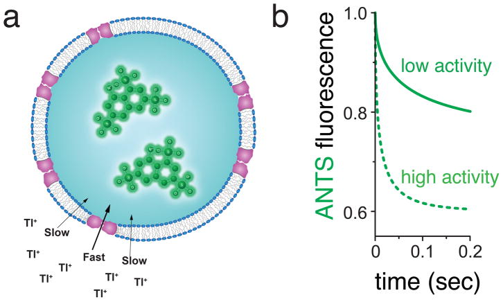
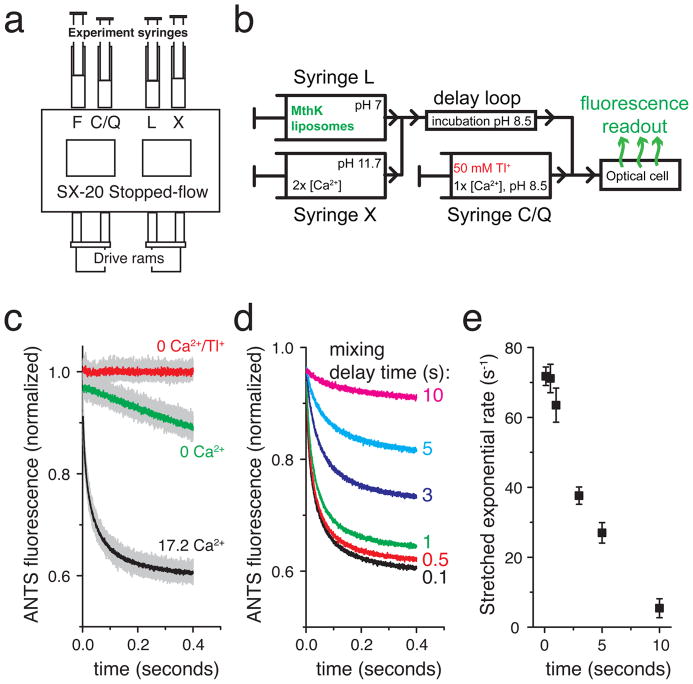
Quantitative investigations into functional properties of purified ion channel proteins using standard electrophysiological methods are challenging, in particular for the determination of average ion channel behavior following rapid changes in experimental conditions (e.g., ligand concentration). Here, we describe a method for determining the functional activity of liposome-reconstituted K+ channels using a stopped-flow fluorometric ion flux assay. Channel activity is quantified by measuring the rate of fluorescence decrease of a liposome-encapsulated fluorophore, specifically quenched by thallium ions entering the liposomes via open channels. This method is well suited for studying the lipid bilayer dependence of channel activity, the activation and desensitization kinetics of ligand-dependent K+ channels, and channel modulation by channel agonists, blockers, or other antagonists.

Keywords: ANTS quenching; Ion channel function; Liposomal ion flux assay; Stopped-flow assay; Thallium.


Figures

Fig. 1
Fig. 1
General principle underlying the detection of K+ channel activity using Tl+ flux measurements. (a) K+ channels are reconstituted into liposomes with ANTS fluorophore inside, and rapidly mixed with Tl+-containing solutions using a stopped-flow mixing device. When channels are open, external Tl+ enter and exchange for K+ exiting the liposomes. The increasing internal Tl+ concentration creates a time-dependent quenching of the ANTS fluorescence inside the liposomes. (b) Higher K+ channel activity leads to faster ANTS fluorescence quenching rate than lower K+ channel activity (dotted line compared to solid line).
Fig. 2
Fig. 2
Kinetic measurements of channel activity are performed using a sequential-mixing stopped-flow spectrofluorometer. (a) Cartoon of the sample handling chamber of the SX20 stopped-flow device (Applied Photophysics). Liposomes in syringe L are mixed with experimental test conditions in syringe X. Following a user-defined reaction delay time, the L+X mixture is further mixed with the contents of syringe C/Q; C denotes a non-quenching control experiment and Q denotes a Tl+-containing quenching experiment. (b) Schematic flow diagram for the sequential mixing steps performed inside the stopped-flow apparatus for an experiment testing the Ca2+ activation of the MthK K+ channel. (c–e) Example data obtained with the MthK Ca2+-activated K+ channel. (c) Raw data from repeated mixing reactions (light gray data) with mean signal (dark data overlay) using 100 ms delay between mixing 1 and 2. Red: 0 Ca2+ in syringe X and 0 Tl+ in syringe C, Green: 0 Ca2+ in syringe X and 50 mM Tl+ in syringe Q, Black: 34.4 mM Ca2+ in syringe X and 50 mM Tl+ in syringe Q. (d) Mean ANTS fluorescence quenching time courses from the experiment in b with the mixing delay time varied between 0.1 and 10 seconds, illustrating the loss of MthK activity upon sustained exposure to Ca2+. (e) Mean stretched exponential rates from fitting individual mixing repeats from c–d to Equation 1 and calculating the rates using Equation 2. The error bars are standard deviations from the repeated mixing reactions as in c. Experimental replicates on independent samples should also be acquired.

References

    1. Aidley DJ. The physiology of excitable cells. 4. Cambridge University Press; Cambridge, UK ; New York, NY, USA: 1998.
    1. Hille B. Ion channels of excitable membranes. 3. Sinauer; Sunderland, Mass: 2001.
    1. Martinac B, Saimi Y, Kung C. Ion channels in microbes. Physiol Rev. 2008;88(4):1449–1490. doi: 10.1152/physrev.00005.2008. - DOI - PMC - PubMed
    1. Cordero-Morales JF, Cuello LG, Zhao Y, Jogini V, Cortes DM, Roux B, Perozo E. Molecular determinants of gating at the potassium-channel selectivity filter. Nature structural & molecular biology. 2006;13(4):311–318. doi: 10.1038/nsmb1069. - DOI - PubMed
    1. Moore HP, Raftery MA. Direct spectroscopic studies of cation translocation by Torpedo acetylcholine receptor on a time scale of physiological relevance. Proceedings of the National Academy of Sciences of the United States of America. 1980;77(8):4509–4513. - PMC - PubMed

Publication types

LinkOut - more resources