

Multichannel wearable fNIRS-EEG system for long-term clinical monitoring
- PMID: 29058341
- PMCID: PMC6866376
- DOI: 10.1002/hbm.23849
Multichannel wearable fNIRS-EEG system for long-term clinical monitoring
Abstract
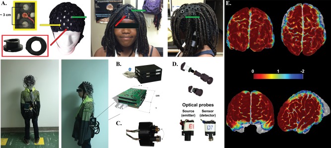
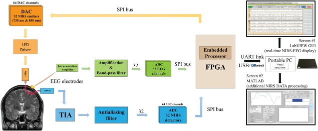
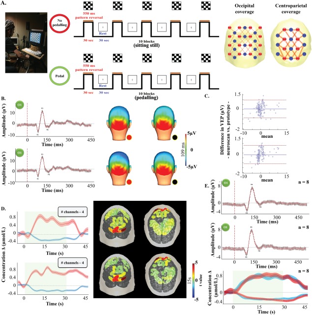
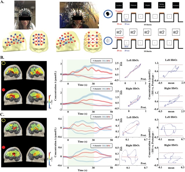
Continuous brain imaging techniques can be beneficial for the monitoring of neurological pathologies (such as epilepsy or stroke) and neuroimaging protocols involving movement. Among existing ones, functional near-infrared spectroscopy (fNIRS) and electroencephalography (EEG) have the advantage of being noninvasive, nonobstructive, inexpensive, yield portable solutions, and offer complementary monitoring of electrical and local hemodynamic activities. This article presents a novel system with 128 fNIRS channels and 32 EEG channels with the potential to cover a larger fraction of the adult superficial cortex than earlier works, is integrated with 32 EEG channels, is light and battery-powered to improve portability, and can transmit data wirelessly to an interface for real-time display of electrical and hemodynamic activities. A novel fNIRS-EEG stretchable cap, two analog channels for auxiliary data (e.g., electrocardiogram), eight digital triggers for event-related protocols and an internal accelerometer for movement artifacts removal contribute to improve data acquisition quality. The system can run continuously for 24 h. Following instrumentation validation and reliability on a solid phantom, performance was evaluated on (1) 12 healthy participants during either a visual (checkerboard) task at rest or while pedalling on a stationary bicycle or a cognitive (language) task and (2) 4 patients admitted either to the epilepsy (n = 3) or stroke (n = 1) units. Data analysis confirmed expected hemodynamic variations during validation recordings and useful clinical information during in-hospital testing. To the best of our knowledge, this is the first demonstration of a wearable wireless multichannel fNIRS-EEG monitoring system in patients with neurological conditions. Hum Brain Mapp 39:7-23, 2018. © 2017 Wiley Periodicals, Inc.
Keywords: cerebral hemodynamics; electroencephalography; epilepsy; functional brain imaging and monitoring; limb-shaking transient ischemic attacks; portable near-infrared spectroscopy.
© 2017 Wiley Periodicals, Inc.
Figures
References
-
- Abtahi M, Cay G, Saikia MJ, Mankodiya K (2016): Designing and testing a wearable, wireless fNIRS patch. Conf Proc IEEE Eng Med Biol Soc 2016: Aug; 2016:6298–6301. - PubMed
-
- Arenth PM, Ricker JH, Schultheis MT (2007): Applications of functional near‐infrared spectroscopy (fNIRS) to neurorehabilitation of cognitive disabilities. Clin Neuropsychol 21:38–57. - PubMed
-
- Atsumori H, Kiguchi M, Obata A, Sato H, Katura T, Utsugi K, Funane T, Maki A (2007): Development of a multi‐channel, portable optical topography system. Conf Proc IEEE Eng Med Biol Soc 2007:3362–3364. - PubMed
Publication types
MeSH terms
Grants and funding
LinkOut - more resources
Full Text Sources
Other Literature Sources
Miscellaneous
