. 2018 Jan;39(1):407-427.
doi: 10.1002/hbm.23852. Epub 2017 Oct 23.

Radiation-induced brain structural and functional abnormalities in presymptomatic phase and outcome prediction

Affiliations

Radiation-induced brain structural and functional abnormalities in presymptomatic phase and outcome prediction

Zhongxiang Ding et al. Hum Brain Mapp. 2018 Jan.

Abstract

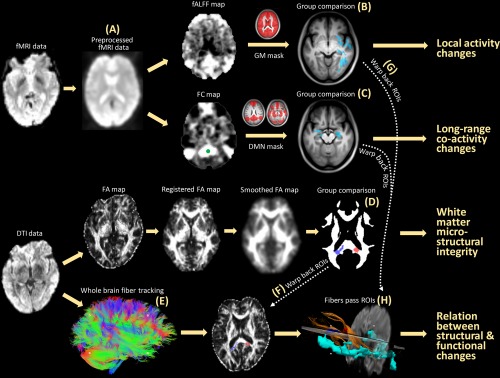
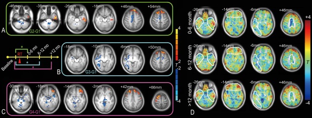
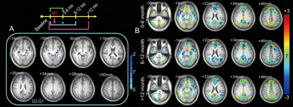
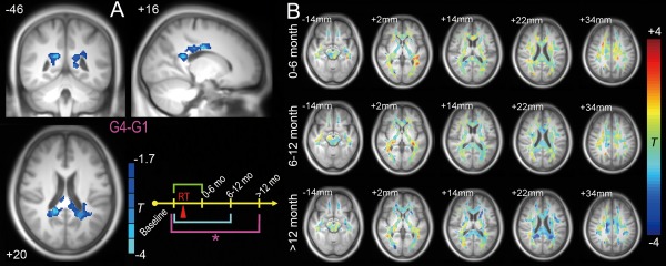
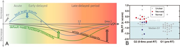
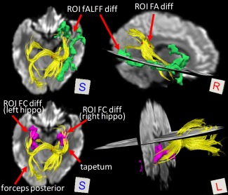
Radiation therapy, a major method of treatment for brain cancer, may cause severe brain injuries after many years. We used a rare and unique cohort of nasopharyngeal carcinoma patients with normal-appearing brains to study possible early irradiation injury in its presymptomatic phase before severe, irreversible necrosis happens. The aim is to detect any structural or functional imaging biomarker that is sensitive to early irradiation injury, and to understand the recovery and progression of irradiation injury that can shed light on outcome prediction for early clinical intervention. We found an acute increase in local brain activity that is followed by extensive reductions in such activity in the temporal lobe and significant loss of functional connectivity in a distributed, large-scale, high-level cognitive function-related brain network. Intriguingly, these radiosensitive functional alterations were found to be fully or partially recoverable. In contrast, progressive late disruptions to the integrity of the related far-end white matter structure began to be significant after one year. Importantly, early increased local brain functional activity was predictive of severe later temporal lobe necrosis. Based on these findings, we proposed a dynamic, multifactorial model for radiation injury and another preventive model for timely clinical intervention. Hum Brain Mapp 39:407-427, 2018. © 2017 Wiley Periodicals, Inc.

Keywords: amplitude of low-frequency fluctuations; diffusion tensor imaging; functional connectivity; functional magnetic resonance imaging; irradiation injury; nasopharyngeal carcinoma; prognosis; radiation therapy; resting state; structural connectivity.


Figures

Figure 1
Figure 1
Schematic flowchart of this study. With fMRI data (A), both the fALFF (measuring local activity) and the seed correlation‐based FC (measuring inter‐regional co‐activity) were calculated and compared between G1 and the other groups (B). With DTI data, a voxel‐wise comparison of the FA (measuring white matter microstructural integrity) was conducted between G1 and the other groups (D). Fiber tracking (E) was performed to link local functional changes (G) and inter‐regional functional integration changes (C) with white matter changes (F, H). [Color figure can be viewed at http://wileyonlinelibrary.com]
Figure 2
Figure 2
Local brain activity changes after RT. Two‐sample t tests were conducted comparing the fALFF before RT (G1, baseline group) with that at different stages after RT (G2: 0–6 months, G3: 6–12 months, G4: >12 months after completion of RT) within a grey matter mask (voxel‐wise t > 2, AlphaSim corrected P < 0.05, cluster size > 172) (A–C). The result was rendered on the averaged T1 images across all subjects. The results from G2 to G1, G3 to G1, and G4 to G1 are framed with green, cyan, and purple, respectively, corresponding to the schematic plot; * indicates that a statistically significant difference was found between groups. The group comparison results are also shown without the threshold (D). The regions with abnormal local activities that changed over time are marked. The brain slices are shown in radiological convention (left is right, right is left). Details of the results are summarized in Table 3. [Color figure can be viewed at http://wileyonlinelibrary.com]
Figure 3
Figure 3
RT‐induced DMN functional connectivity changes. Only G3 G1 (6–12 months after RT vs pre‐RT) revealed a significant FC reduction within the DMN (t > 2.02, P < 0.05, cluster size > 101, corrected by AlphaSim) (A). The unthresholded results for each post‐RT stage on typical slices of the DMN are also shown (B), with a significant whole network FC reduction at 6–12 months after RT. The results were rendered on the averaged T1 images across all subjects. The brain slices are shown in radiological convention (left is right, right is left). Details of the results are summarized in Table 4. [Color figure can be viewed at http://wileyonlinelibrary.com]
Figure 4
Figure 4
FA reductions 12 months or longer from the completion of RT. Only G4 G1 reached statistical significance (Gaussian Random Field‐based correction, cluster‐wise P < 0.05, voxel‐wise P < 0.05, single‐tailed statistical test as implemented in SPM8) (A). FA changes compared with G1 (pre‐RT) at all three stages are shown without thresholds in several typical slices. The underlying image is the averaged T1 image across all subjects. The brain slices are shown in radiological convention (left is right, right is left). Details are summarized in Table 4. [Color figure can be viewed at http://wileyonlinelibrary.com]
Figure 5
Figure 5
Hypothesized model of different recovery/progression courses after RT with and without early intervention (A) and potential predictive biomarkers with early fALFF increments (B). In our hypothesized recovery/progressive model, a local temporal lobe brain activity increment preceded later default‐mode FC and even later white matter integrity reductions. This model is plotted by the solid curves. We also proposed a preventive model, shown by the dotted curves; in this model, if the earlier local brain activity changes could be continuously monitored and carefully controlled by neuroprotective treatment, severe later white matter integrity loss and temporal lobe necrosis might be prevented. In (B), the early abnormal fALFF increment was plotted with the later outcomes (necrosis, indicating bad outcomes; or no necrosis after 5 years, indicating good outcomes) to evaluate the prognostic value of our proposed early biomarker and to further validate our preventive model. Specifically, based on the peak coordinates of G2 G1 group differences in fALFF in the left inferior temporal lobe ( 45 9 39, see Table 3), a sphere ROI was generated (radius = 6 mm). Mean fALFF (in z score) across voxels in this ROI was extracted for each subject in G1 (pre‐RT) and G2 (0–6 months after RT). Subjects showing clear temporal lobe necrosis during follow‐up are shown in red; those whose brains still appeared normal are shown in blue. Subjects with unclear outcomes due to dropout are shown in gray. The median fALFF value for G2 subjects (z = −0.44) delineates a warning line that appears as a borderline between the light blue (safe) and the white (dangerous) zones and clearly separates the two groups of subjects with different outcomes. The median fALFF values for G1 subjects are shown by a dashed line. [Color figure can be viewed at http://wileyonlinelibrary.com]
Figure 6
Figure 6
Fibers connect regions showing earlier functional and later structural changes. The result shown is derived from a randomly selected subject. Fiber bundles (yellow streamlines) were found to link earlier local activity changes in the far‐end temporal cortex (green clusters) and earlier default mode FC changes in the bilateral hippocampus (purple clusters) and subsequent late‐delayed white‐matter microstructural integrity changes in the right‐side splenium of the corpus callosum (red cluster). [Color figure can be viewed at http://wileyonlinelibrary.com]
Figure 7
Figure 7
Correlations between the fALFF and FC values in the hippocampus (A) and between the FC and the irradiation dose (B). An intersecting ROI was generated by overlapping the fALFF reduction and FC reduction results in the G3 G1 comparison centered at the MNI coordinates of 27 24 6 in the right hippocampus. The mean fALFF and FC values within the ROI were extracted for all subjects from all groups and plotted. The FC with the PCC and the regional fALFF correlated with one another across all subjects in all groups (r = 0.23, P = 0.03). Two ROIs in the bilateral hippocampus that showed a significantly reduced FC with the PCC in the G3 G1 comparison were selected for dose–relationship analysis. The ROIs consisted of two spheres centered at the peak MNI coordinates 30 15 15 (right hippocampus) and 18 18 3 (left hippocampus) with a 6 mm radius. The averaged FC on each side was correlated with the maximum and mean RT doses to the corresponding side of the temporal lobe across nine subjects who received IMRT and in whom the dose could be calculated. The maximum irradiation doses in the right temporal lobe correlated with the PCC‐to‐right hippocampus FC (r = −0.66, P < 0.05). [Color figure can be viewed at http://wileyonlinelibrary.com]

References

    1. Abayomi OK (1996): Pathogenesis of irradiation‐induced cognitive dysfunction. Acta Oncologica 35:659–663. - PubMed
    1. Acker JC, Marks LB, Spencer DP, Yang W, Avery MA, Dodge RK, Rosner GL, Dewhirst MW (1998): Serial in vivo observations of cerebral vasculature after treatment with a large single fraction of radiation. Radiation Research 149:350–359. - PubMed
    1. Akiyama K, Tanaka R, Sato M, Takeda N (2001): Cognitive dysfunction and histological findings in adult rats one year after whole brain irradiation. Neurologia medico‐Chirurgica 41:590–598. - PubMed
    1. Atwood T, Payne VS, Zhao W, Brown WR, Wheeler KT, Zhu JM, Robbins ME (2007): Quantitative magnetic resonance spectroscopy reveals a potential relationship between radiation‐induced changes in rat brain metabolites and cognitive impairment. Radiation Research 168:574–581. - PubMed
    1. Biswal BB (2012): Resting state fMRI: A personal history. NeuroImage 62:938–944. - PubMed

MeSH terms

LinkOut - more resources