. 2017 Dec 1;127(12):4242-4256.
doi: 10.1172/JCI92309. Epub 2017 Oct 23.

Endothelial jagged-2 sustains hematopoietic stem and progenitor reconstitution after myelosuppression

Affiliations

Endothelial jagged-2 sustains hematopoietic stem and progenitor reconstitution after myelosuppression

Peipei Guo et al. J Clin Invest. .

Abstract

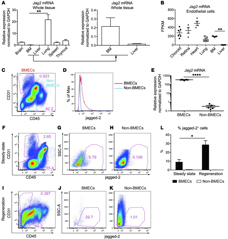
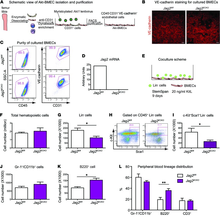
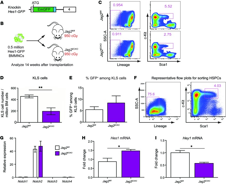
Angiocrine factors, such as Notch ligands, supplied by the specialized endothelial cells (ECs) within the bone marrow and splenic vascular niche play an essential role in modulating the physiology of adult hematopoietic stem and progenitor cells (HSPCs). However, the relative contribution of various Notch ligands, specifically jagged-2, to the homeostasis of HSPCs is unknown. Here, we show that under steady state, jagged-2 is differentially expressed in tissue-specific vascular beds, but its expression is induced in hematopoietic vascular niches after myelosuppressive injury. We used mice with EC-specific deletion of the gene encoding jagged-2 (Jag2) to demonstrate that while EC-derived jagged-2 was dispensable for maintaining the capacity of HSPCs to repopulate under steady-state conditions, by activating Notch2 it did contribute to the recovery of HSPCs in response to myelosuppressive conditions. Engraftment and/or expansion of HSPCs was dependent on the expression of endothelial-derived jagged-2 following myeloablation. Additionally, jagged-2 expressed in bone marrow ECs regulated HSPC cell cycle and quiescence during regeneration. Endothelial-deployed jagged-2 triggered Notch2/Hey1, while tempering Notch2/Hes1 signaling in HSPCs. Collectively, these data demonstrate that EC-derived jagged-2 activates Notch2 signaling in HSPCs to promote hematopoietic recovery and has potential as a therapeutic target to accelerate balanced hematopoietic reconstitution after myelosuppression.

Keywords: Adult stem cells; Bone marrow; Hematology; Vascular Biology; endothelial cells.


Conflict of interest statement

Conflict of interest: The authors have declared that no conflict of interest exists.

Figures

Figure 1
Figure 1. Jagged-2 is dynamically expressed in BMECs.
(A) The expression level of Jag2 mRNA in different mouse whole organs (n = 3). The mRNA expression is calculated using GAPDH as internal control. (B) The FPKM (fragments per kilobase of exon per million fragments mapped) value for Jag2 mRNA in primary ECs from various organs. The number of dots indicates the number of biological replicates. (C) Representative flow cytometric plots for the gating of CD31+CD45 BMECs and CD31CD45 non-BMECs (n = 4). (D) Histogram of jagged-2 expression on BMECs and non-BMECs. (E) qPCR quantification of Jag2 mRNA from sorted BMECs (n = 3) and non-BMECs (n = 5). The RNA expression level is calculated using GAPDH as internal control. (FH) Representative flow plots for jagged-2 expression in BMECs and non-BMECs (n = 4) under homeostatic conditions. (IK) Jagged-2 expression within BMECs and non-BMECs at 2 weeks after 650 cGy sublethal irradiation (n = 5). (L) Comparison of percentage of jagged-2+ cells among BMECs under steady state and during regeneration after myeloablative injuries. Error bars indicate the SEM. *P < 0.05, **P < 0.01, and ****P < 0.0001, by 2-tailed unpaired t test. The numbers in the flow plots represent percentages of cells.
Figure 2
Figure 2. Endothelial-supplied jagged-2 is not required to maintain HSPC function under homeostatic conditions.
(AD) The total number of BMMNCs (A) and the WBC count (C) were quantified in adult Jag2fl/fl or Jag2ECKO mice. Lineage distribution of hematopoietic cells within the BM (B) and peripheral blood (D) was quantified in Jag2fl/fl or Jag2ECKO mice. For A and B, n = 3. For C, the number of dots indicates the number of biological replicates. For D, n = 9 for Jag2fl/fl, n = 8 for Jag2ECKO. (E) Representative flow cytometric gating of phenotypic Linc-Kit+Sca1+CD150+CD48 long-term HSCs (LT-HSCs). (F) Quantification of the number of phenotypic LT-HSCs per million BMMNCs in the femur of Jag2fl/fl or Jag2ECKO (n = 8 for each group). (G) Schematic view of competitive repopulating assay. (H) The percentage of CD45.2+ hematopoietic cells in the peripheral blood of CD45.1+ mice at 15.5 weeks after transplantation (n = 6 CD45.1 recipients for Jag2fl/fl and n = 8 CD45.1 recipients for Jag2ECKO mice). (I) Representative flow cytometric plots showing the multilineage engraftment. The CD45.2+ cells were further gated to reveal donor-derived CD3+ T cells and CD19+ B cells. (J) Quantification of multilineage engraftment within the CD45.2+ cells. Error bars indicate the SEM. *P < 0.05 and **P < 0.01, by unpaired 2-tailed t test. The numbers in the flow plots represent percentages of cells.
Figure 3
Figure 3. Jagged-2 expressed by BMECs promotes in vitro expansion of HSPCs.
(A) Schematic view of the method to enrich BMECs from Jag2fl/fl or Jag2ECKO mice. (B) The cultured BMECs were stained with anti–VE-cadherin antibody. Scale bars: 100 μm. (C) After passages, the purity of BMECs from Jag2fl/fl or Jag2ECKO mice was confirmed by flow cytometric staining of CD45, CD31, and VE-cadherin (n = 5). The percentage of CD45CD31+VE-cadherin+ ECs is shown. (D) Using primers flanking the first 2 exons of Jag2 mRNA, the expression level of Jag2 was quantified via real-time qPCR (n = 3). (E) Schematic view of the coculture setup using BMECs and lineage-negative (Lin) hematopoietic cells (n = 3 biological replicates of Lin cells were used for the coculture; 1 Jag2fl/fl BMEC and 2 lines of Jag2ECKO BMECs were used as feeders). (FI) At day 9 after coculture, the total number of CD45+ cells (F), Lin cells (G), and KLS cells (I) within the culture was summarized. The representative flow cytometric plots of KLS HSPCs are shown in H. (J and K) At day 9 after coculture, the total number of Gr-1+/CD11b+ myeloid cells (J) and B220+ cells (K) was quantified. (L) Quantification of peripheral blood multilineage distribution in 18-month-old Jag2fl/fl or Jag2ECKO mice (n = 7 for Jag2fl/fl, n = 8 for Jag2ECKO). Error bars indicate the SEM. *P < 0.05 and **P < 0.01, by 2-tailed t test. The numbers in the flow plots represent percentages of cells.
Figure 4
Figure 4. Endothelial jagged-2 ensures proper HSPC recovery after myelosuppression.
(A) Jag2fl/fl or Jag2ECKO mice were subjected to weekly 5-fluorouracil (5-FU) injections at the dose of 150 mg/kg. Kaplan-Meier survival curve was generated after monitoring of the survival rate of the mice. n = 12 for Jag2fl/fl, n = 9 for Jag2ECKO mice. (B) Jag2fl/fl or Jag2ECKO mice were subjected to sublethal irradiation at 650 cGy, and their survival rate was monitored. This experiment was performed 4 times. Each time, n = 4 mice were used for each genotype. The data are combined and shown in B. (CH) To monitor the kinetics of hematopoiesis regeneration following irradiation, the quantification of KLS cells and LT-HSCs at day 10 (C and D) (n = 4 for each genotype), day 16 (E and F) (n = 5 for each group), and day 29 (G and H) (n = 5 for each group) after sublethal irradiation is further shown. Error bars indicate the SEM. *P < 0.05 and **P < 0.01, by unpaired 2-tailed t test.
Figure 5
Figure 5. Vascular perfusion function is preserved in Jag2ECKO mice following myelosuppression.
At day 10 after 650 cGy irradiation, the perfusion function of spleen vasculature (A), liver vasculature (B), BM vasculature (C), and lung vasculature (D) was preserved in Jag2ECKO mice, as demonstrated by fluorescence-labeled VE-cadherin antibodies perfused into the vasculature (n = 4 for each group). Scale bars: 50 μm.
Figure 6
Figure 6. Endothelial jagged-2 modulates the cell-cycle status of HSPCs during regeneration.
(A) On day 16 after irradiation, the flow cytometric gating for KLS, CD150CD48 short-term KLS (ST-HSCs), and CD150CD48+ KLS multipotent progenitor cells is shown. (B) At day 16 after irradiation, the percentage of Ki67 G0 cells among KLS cells was quantified (n = 5 for each group). (C) At day 14 after irradiation, the percentage of apoptotic cells among Lin cells, cKit+Lin cells, KLS cells, and Sca1+Lin cells was quantified. (D) On day 29 after 650 cGy irradiation, the percentage of Ki67 G0 cells among Lin cells and KLS cells was quantified (n = 5 for each group). Error bars indicate the SEM. *P < 0.05, by unpaired 2-tailed t test. The numbers in the flow plots represent percentages of cells.
Figure 7
Figure 7. Endothelial jagged-2 induces Notch2/Hey1 signaling in HSPCs, enhancing engraftment.
(A and B) Schematic view of the knockin Hes1-GFP reporter mouse line (A) and the transplantation assays (B) used to test the role of BMEC jagged-2 in promoting engraftment/expansion of HSPCs. n = 4 for Jag2fl/fl group, n = 3 for Jag2ECKO group. (C) Representative flow plots for the gating strategies for KLS cells in the Jag2fl/fl and Jag2ECKO mice. (D) The number of HSPCs per million BM cells was quantified. (E) The percentage of GFP+ cells among the gated KLS cells is summarized. The data were collected following the transplantation experiment listed in B. (F) Representative flow cytometric plots showing the sorting strategies for KLS cells from Jag2fl/fl or Jag2ECKO mice. (GI) Real-time qPCR analysis of the sorted HSPCs from Jag2fl/fl or Jag2ECKO mice was carried out for Notch receptors (G) and Notch targets (H and I). For G and I, n = 3 biological replicates for each group. For H, n = 5 for each group. Error bars indicate the SEM. *P < 0.05 and **P < 0.01, by unpaired 2-tailed t test. The numbers in the flow plots represent percentages of cells.
Figure 8
Figure 8. Jagged-2 supplied by ECs and hematopoietic cells maintains HSC number.
(A) Summary of mRNA expression of Notch ligands Jag1, Jag2, Dll1, Dll3, and Dll4 in HSPCs. Data are from the published RNA sequencing data of SP-KLS-CD150+ cells (29). VE-cadherin-Cre mice (30) were crossed with Jag2fl/fl mice to delete exons 1 and 2 of the Jag2 gene from ECs and hematopoietic cells. (B and C) After crossing of VE-cadherin-Cre mice with Rosa26CAG<stop>tdtomato mice, the deletion efficiency of VE-cadherin-Cre in ECs (B) and hematopoietic cells within the BM (C) was quantified. (DF) The platelets (D), WBC (E), and RBC (F) were monitored after sublethal irradiation with 650 cGy at the indicated time points. (G) The survival curve of Jag2fl/fl or Jag2KO mice was plotted. (H and I) Competitive repopulating assay was carried out using BMMNCs from Jag2fl/fl or Jag2KO mice (n = 7 for each group). For B and C, n = 3 biological replicates for each group. For DG, n = 5 for Jag2fl/fl, n = 4 for Jag2KO mice. For I, n = 7 each for Jag2fl/fl and Jag2KO mice. Error bars indicate SEM. For DF, at each individual time point, the difference between Jag2fl/fl and Jag2KO mice was compared using a 2-tailed t test. The resulting P value is shown. For DF and J, the overall differences of the 2 curves were also compared using 2-way ANOVA, and the P values of the observed variance based on the genotype are as follows: for D, P = 0.0025; for E, P < 0.0001; for F, P = 0.0373; for I, P = 0.0016. *P < 0.05; **P < 0.01 (**P value was determined by 2-way ANOVA).

References

    1. Mendelson A, Frenette PS. Hematopoietic stem cell niche maintenance during homeostasis and regeneration. Nat Med. 2014;20(8):833–846. doi: 10.1038/nm.3647. - DOI - PMC - PubMed
    1. Ramalingam P, Poulos MG, Butler JM. Regulation of the hematopoietic stem cell lifecycle by the endothelial niche. Curr Opin Hematol. 2017;24(4):289–299. doi: 10.1097/MOH.0000000000000350. - DOI - PMC - PubMed
    1. Poulos MG, et al. Endothelial Jagged-1 is necessary for homeostatic and regenerative hematopoiesis. Cell Rep. 2013;4(5):1022–1034. doi: 10.1016/j.celrep.2013.07.048. - DOI - PMC - PubMed
    1. Heissig B, et al. Recruitment of stem and progenitor cells from the bone marrow niche requires MMP-9 mediated release of kit-ligand. Cell. 2002;109(5):625–637. doi: 10.1016/S0092-8674(02)00754-7. - DOI - PMC - PubMed
    1. Rafii S, Butler JM, Ding BS. Angiocrine functions of organ-specific endothelial cells. Nature. 2016;529(7586):316–325. doi: 10.1038/nature17040. - DOI - PMC - PubMed

Publication types