. 2017 Nov;19(11):1326-1335.
doi: 10.1038/ncb3632. Epub 2017 Oct 23.

Synthetic hydrogels for human intestinal organoid generation and colonic wound repair

Affiliations

Synthetic hydrogels for human intestinal organoid generation and colonic wound repair

Ricardo Cruz-Acuña et al. Nat Cell Biol. 2017 Nov.

Abstract

In vitro differentiation of human intestinal organoids (HIOs) from pluripotent stem cells is an unparalleled system for creating complex, multicellular three-dimensional structures capable of giving rise to tissue analogous to native human tissue. Current methods for generating HIOs rely on growth in an undefined tumour-derived extracellular matrix (ECM), which severely limits the use of organoid technologies for regenerative and translational medicine. Here, we developed a fully defined, synthetic hydrogel based on a four-armed, maleimide-terminated poly(ethylene glycol) macromer that supports robust and highly reproducible in vitro growth and expansion of HIOs, such that three-dimensional structures are never embedded in tumour-derived ECM. We also demonstrate that the hydrogel serves as an injection vehicle that can be delivered into injured intestinal mucosa resulting in HIO engraftment and improved colonic wound repair. Together, these studies show proof-of-concept that HIOs may be used therapeutically to treat intestinal injury.


Figures

Figure 1
Figure 1. PEG-4MAL polymer density and adhesive ligand type control HIO viability
(a) Transmitted light and fluorescence microscopy images of HIOs cultured in PEG-4MAL hydrogels of different polymer density or Matrigel™. HIO viability was assessed at 7 d after encapsulation. Bar, 500 μm. (b) Percentage of total organoid area stained for live or dead (mean ± SEM) after 7 d of encapsulation (n = 5 organoids analyzed for all groups, except n = 6 organoids analyzed for 4.0% group). (c) Transmitted light and fluorescence microscopy images of HIOs cultured in 4.0% PEG-4MAL hydrogels functionalized with different adhesive peptides or Matrigel™. HIO viability was assessed at 7 d after encapsulation. Bar, 500 μm. (d) Percentage of total organoid area stained for live or dead (mean ± SEM) after 7 d of encapsulation (n = 5 organoids analyzed for all groups, except n = 6 organoids analyzed for AG73 and Matrigel groups). (b,d) One-way ANOVA with Tukey's multiple comparisons test showed significant differences between (b) 4.0% PEG-4MAL or Matrigel™ and 5.0 or 6.0% PEG-4MAL, and between (d) PEG-4MAL-RGD or Matrigel™ and PEG-4MAL-GFOGER or -IKVAV. (*P < 0.05, **P < 0.01, ***P < 0.001, ****P < 0.0001). (a-d) Three independent experiments were performed and data is presented for one of the experiments. Every independent experiment was performed with 6 gel samples per experimental group (PEG-4MAL, Matrigel™). Source data are available in Supplementary Table 1.
Figure 2
Figure 2. Engineered PEG-4MAL supports HIO development
(a) Transmitted light and fluorescence microscopy images of HIOs cultured in 4.0% PEG-4MAL hydrogels functionalized with (a) RGD, (b) inactive, scrambled RDG peptide, or (c) non-degradable crosslinker (DTT). HIO viability was assessed at 7 d after encapsulation. (d,e) Transmitted light microscopy images of Matrigel™-generated HIOs cultured within (d) 4.0% PEG-4MAL-RGD hydrogel or (e) Matrigel™ over time. Bar, 500 μm. (f,g) Fluorescence microscopy images of a HIO at 7 d after encapsulation in 4.0% PEG-4MAL-RGD hydrogel or Matrigel™, and labeled for (f) β-CATENIN, proliferative cells (KI67), and (g) epithelial apical polarity (EZRIN) and tight junctions (ZO-1). DAPI, counterstain. “L” indicates HIO lumen. Bar, 100 μm. (a-g) Three independent experiments were performed and data is presented for one of the experiments. Every experiment was performed with (a-c) 4, (f,g) 6 or (d,e) 12 gel samples per experimental group (PEG-4MAL, Matrigel™).
Figure 3
Figure 3. PEG-4MAL polymer density regulates HIO generation from intestinal spheroids in the absence of Matrigel
embedding. Transmitted light and fluorescence microscopy images of mCherry-spheroids cultured in PEG-4MAL hydrogels of different polymer density or Matrigel™. Spheroids viability was assessed by Calcein-AM labeling at 2 h after encapsulation (day 0) and at day 5 for PEG-4MAL conditions, and at day 3 for Matrigel™. Bar, 100 μm. Viability is quantified as percentage of total spheroid or organoid area stained for live or dead (mean ± SEM; n = 5 organoids analyzed per condition/time-point). One-way ANOVA with Tukey's multiple comparisons test showed significant differences between 3.5% or 4.0% PEG-4MAL and 8.0% or 12.0% PEG-4MAL at Day 0 (****P < 0.0001). (b) HIO projected area and (c) Feret diameter normalized to Day 0 values at different time-points after encapsulation in 4.0% PEG-4MAL-RGD hydrogel ( formula image
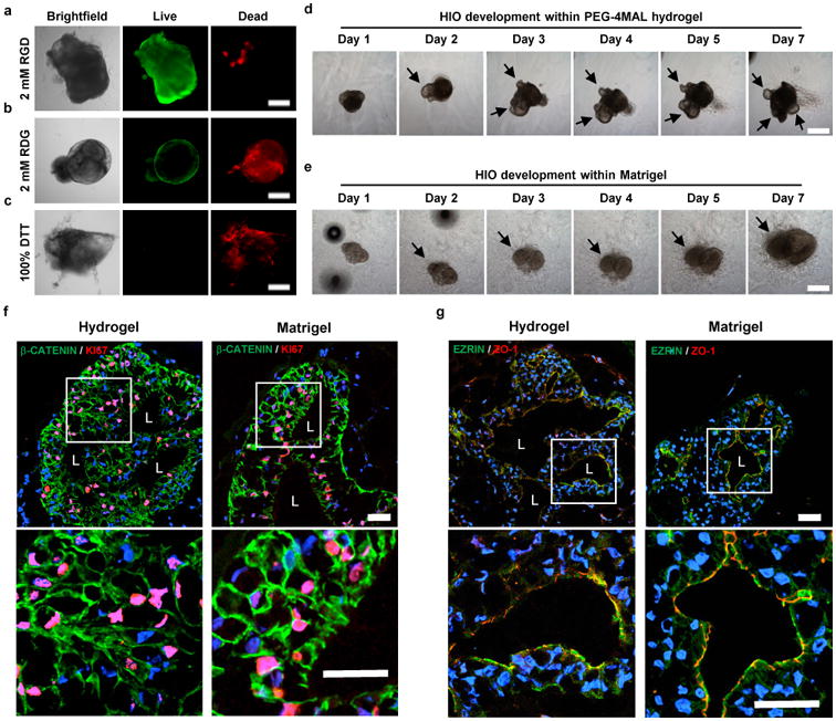
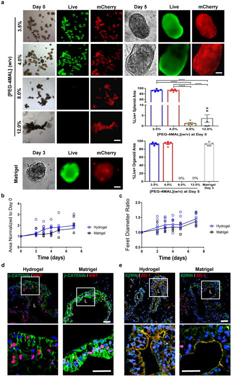
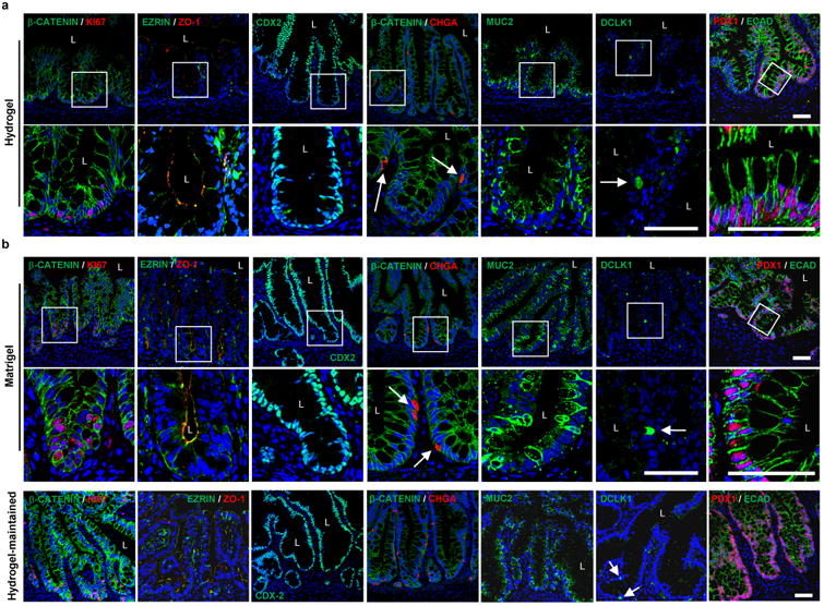
) or Matrigel™ (■) (n = 6 organoids for PEG-4MAL and n = 4 organoids for Matrigel™ per time-point). Repeated measures two-way ANOVA showed no significant difference between matrix types (P > 0.05). Line represents the mean of the individual data points at each time-point. (d,e) Fluorescence microscopy images of a HIO at 21 d after encapsulation in 4.0% PEG-4MAL-RGD hydrogel or Matrigel™ and labeled for (d) β-CATENIN, proliferative cells (KI67), and (e) epithelial apical polarity (EZRIN) and tight junctions (ZO-1). DAPI, counterstain. “L” indicates HIO lumen. Bars, 100 μm. Three independent experiments were performed and data is presented for one of the experiments. Every experiment was performed with 6 gel samples per experimental group (PEG-4MAL, Matrigel™). Source data are available in Supplementary Table 1.
Figure 4
Figure 4. PEG-4MAL-generated HIOs develop a mature intestinal tissue structure in vivo
(a) Micrographs of dissected kidneys containing HIOs generated within PEG-4MAL hydrogel, Matrigel™, or generated within Matrigel™ and maintained within PEG-4MAL (hydrogel-maintained). (b) Transmitted light and fluorescence microscopy (mCherry) images of harvested organoids. Bar, 0.5 cm. (c,d) H&E staining demonstrates mature human intestinal crypt-villus structure, and Alcian blue and trichrome staining reveal presence of differentiated goblet cells and organized collagen fibers. Bar, 100 μm. One experiment was performed using 3 mice per experimental group.
Figure 5
Figure 5. PEG-4MAL-generated HIOs differentiate into mature intestinal tissue in vivo
(a,b) Fluorescence microscopy images of HIOs generated within (a) PEG-4MAL hydrogel, (b) Matrigel™, or generated within Matrigel™ and maintained within PEG-4MAL (hydrogel-maintained), and labeled for β-CATENIN, proliferative cells (KI67), epithelial apical polarity (EZRIN) and junctions (ZO-1 and ECAD), intestinal epithelial protein CDX2, enteroendocrine cells (CHGA), goblet cells (MUC2), tuft cells (DCLK1) and small intestinal marker (duodenum; PDX1). DAPI, counterstain. “L” indicates HIO lumen. White arrows show enteroendocrine cells or tuft cells. Bars, 50 μm. One experiment was performed using 3 mice per experimental group.
Figure 6
Figure 6. PEG-4MAL serves as an injectable delivery vehicle to promote HIO engraftment and wound closure
(a) PEG-4MAL-generated HIOs mixed with engineered hydrogel precursor solutions were injected underneath mechanically-induced mucosal wounds, as seen through the colonoscope camera. (b) Mechanically-induced mucosal wound and fluorescence imaging (mCherry) at the wound site at 5 d post-injection. Bar, 500 μm. (c) Fluorescence microscopy images of murine colonic tissue at the wound site labeled for human cell nuclei (NUMA) at 4 weeks post-delivery. Left: Images from wound edge showing insets from i) adjacent host tissue and ii) wound. Right: Images from wound center showing insets at wound site. DAPI, counterstain. Bars, 100 μm. (d) In situ hybridization, stained for human OLFM4+ cells. Bar, 50 μm. (e) Images of mucosal wounds at 1 d (prior to injection) or 5 d post-injury in murine colon as seen through the colonoscope camera. Mucosal wound area at 5 d post-injury was normalized to day 1 (prior to injection) values (mean ± SEM). Five colonic wounds per mouse were analyzed and averaged (n = 4 mice per condition). One-way ANOVA with Tukey's multiple comparisons test showed significant difference between hydrogel-grown HIOs in hydrogel ( formula image) or Matrigel™-grown HIOs in hydrogel ( formula image) and saline-injected HIOs ( formula image), only hydrogel ( formula image
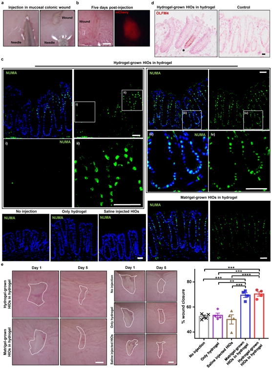
), or no injection group (X) (**P < 0.01, ***P < 0.001, ****P < 0.0001). Bar, 500 μm. Two independent experiments were performed and data is presented for one of the experiments. Experiments performed with 4 mice per experimental group (five colonic wounds/injections per mouse; a-e). Source data are available in Supplementary Table 1.

References

    1. Thomson JA, et al. Embryonic Stem Cell Lines Derived from Human Blastocysts. Science. 1998;282:1145–1147. doi: 10.1126/science.282.5391.1145. - DOI - PubMed
    1. Takahashi K, et al. Induction of Pluripotent Stem Cells from Adult Human Fibroblasts by Defined Factors. Cell. 2007;131:861–872. doi: 10.1016/j.cell.2007.11.019. - DOI - PubMed
    1. Takahashi K, Yamanaka S. A developmental framework for induced pluripotency. Development. 2015;142:3274–3285. doi: 10.1242/dev.114249. - DOI - PubMed
    1. Fox IJ, et al. Stem cell therapy. Use of differentiated pluripotent stem cells as replacement therapy for treating disease. Science. 2014;345:1247391. doi: 10.1126/science.1247391. - DOI - PMC - PubMed
    1. Robinton DA, Daley GQ. The promise of induced pluripotent stem cells in research and therapy. Nature. 2012;481:295–305. doi: 10.1038/nature10761. - DOI - PMC - PubMed