Comparative Study
. 2018 Feb;36(2):699-710.
doi: 10.1002/jor.23779. Epub 2017 Nov 22.

Osteophytes and fracture calluses share developmental milestones and are diminished by unloading

Affiliations
Comparative Study

Osteophytes and fracture calluses share developmental milestones and are diminished by unloading

Allison W Hsia et al. J Orthop Res. 2018 Feb.

Abstract

Osteophytes are a typical radiographic finding during osteoarthritis (OA), but the mechanisms leading to their formation are not well known. Comparatively, fracture calluses have been studied extensively; therefore, drawing comparisons between osteophytes and fracture calluses may lead to a deeper understanding of osteophyte formation. In this study, we compared the time courses of osteophyte and fracture callus formation, and investigated mechanisms contributing to development of these structure. Additionally, we investigated the effect of mechanical unloading on the formation of both fracture calluses and osteophytes. Mice underwent either transverse femoral fracture or non-invasive anterior cruciate ligament rupture. Fracture callus and osteophyte size and ossification were evaluated after 3, 5, 7, 14, 21, or 28 days. Additional mice were subjected to hindlimb unloading after injury for 3, 7, or 14 days. Protease activity and gene expression profiles after injury were evaluated after 3 or 7 days of normal ambulation or hindlimb unloading using in vivo fluorescence reflectance imaging (FRI) and quantitative PCR. We found that fracture callus and osteophyte growth achieved similar developmental milestones, but fracture calluses formed and ossified at earlier time points. Hindlimb unloading ultimately led to a threefold decrease in chondro/osteophyte area, and a twofold decrease in fracture callus area. Unloading was also associated with decreased inflammation and protease activity in injured limbs detected with FRI, particularly following ACL rupture. qPCR analysis revealed disparate cellular responses in fractured femurs and injured joints, suggesting that fracture calluses and osteophytes may form via different inflammatory, anabolic, and catabolic pathways. © 2017 Orthopaedic Research Society. Published by Wiley Periodicals, Inc. J Orthop Res 36:699-710, 2018.

Keywords: fracture healing; hindlimb unloading; osteophytes; post-traumatic osteoarthritis.


Figures

Figure 1
Figure 1. MicroCT analysis of fracture calluses and ACL ruptured joints
(A) Three-dimensional μCT renderings of fracture calluses (left) and osteophytes (right). Callus and osteophyte volume depicted in dark grey, native bone in light grey. (B) Osteophyte and fracture callus volume at days 14, 21, and 28 after injury. Mineralized fracture callus volume remained statistically similar at each time point, while osteophyte volume increased over time (p<0.0001). A significant interaction between time point and injury type indicated differences in trajectory for fracture callus and osteophyte growth (p=0.0375). (C) Osteophyte and fracture callus bone tissue mineral density (TMD) at days 14, 21, and 28 after injury. Fracture callus TMD increased significantly over time, while osteophyte TMD did not change (p=0.0017). Time points that do not share a letter are significantly different from each other (p<0.05). Data is presented as mean ± SD.
Figure 2
Figure 2. Histological assessment of osteophyte and fracture callus size and ossification
(A) Representative H&E stained images for fracture calluses and chondro/osteophytes at 7, 14, 21, and 28 days post-injury (FC= fracture callus; F= femur; M= meniscus; T= tibia). Scale bar is 1 mm. Fracture callus and chondro/osteophyte areas outlined in black. (B) Fracture callus and chondro/osteophyte areas as a function of days after injury. Fracture callus growth occurred at earlier time points, with remodeling and a subsequent decrease in area at later time points. Osteophyte growth occurred at comparatively later time points, but continued throughout the duration of the experiment. A significant interaction between time point and injury type indicates statistical differences in growth trend (p=0.0002). (C) Ossification percentage for fracture calluses and chondro/osteophytes over time. Fracture calluses exhibit earlier ossification, eventually plateauing by days 21–28. Osteophyte ossification continuously increased started at day 14 after injury. Time points that do not share a letter are significantly different from each other (p<0.05). Data is presented as mean ± SD.
Figure 3
Figure 3. Histological comparison of fracture callus and osteophyte composition
Representative H&E stained images for fracture calluses (ground control days 7 and 14, hindlimb unloaded day 7) and osteophytes (days 14 and 21, hindlimb unloaded day 14). Both fracture calluses and osteophytes exhibited similar milestone stages of fracture healing, but at different time points. Fracture calluses exhibited robust chondrogenesis and intramembranous bone formation at day 7, similar to osteophyte composition at day 14. Similarly, fracture calluses underwent endochondral ossification by day 14, while osteophyte endochondral ossification did not occur until day 21. ACL ruptured GC mice exhibited fibrocartilaginous thickening of the synovium and chondrocyte hypertrophy in the meniscus by 14 days post-injury. HLU limbs exhibited decreased inflammation, including reduced periosteal and synovial thickening for fractured and ACL ruptured limbs, respectively, as well as reduced cell infiltration via the periosteum and synovium.
Figure 4
Figure 4. Reduced osteophyte formation in unloaded injured hindlimbs
(A) Representative H&E stained images for ground control (GC) and hindlimb unloaded (HLU) chondro/osteophytes and fracture calluses 7 and 14 days after injury (F = femur, T = tibia, FC = fracture callus). Osteophyte and fracture callus areas outlined in black. Scale bar = 1 mm. (B) HLU reduced osteophyte and fracture callus size. HLU fracture callus areas were not significantly different between time points, but were significantly less than ground control callus areas at each time point (p=0.0077). Similarly, HLU chondro/osteophyte areas were not significantly different at each time point, but were significantly lower than ground control areas at day 14 (p=0.0009). Fracture callus data not connected by the same letter are significantly different. Asterisk indicates that osteophyte area for GC mice at day 14 is significantly greater than all other ACL rupture groups. (C) HLU affected ossification percentage in fracture calluses, but not osteophytes. HLU fracture calluses were significantly lower in ossification percentage at day 7 compared to GC calluses, but the two groups were not significantly different by day 14 (p=0.0042). Conversely, HLU did not affect ossification percentage for osteophytes. Fracture callus data not connected by the same letter are significantly different. # indicates that osteophyte ossification percentages varied by time point (p<0.0001). Both area and ossification percentage were significantly different depending on injury type (p<0.0001). Data is presented as mean ± SD.
Figure 5
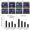
Figure 5. Decreased inflammatory protease activity in unloaded injured limbs
(A) Representative FRI images of ground control (GC) and hindlimb unloaded (HLU) limbs at days 3 and 7 after fracture or ACL rupture. Pink circles represent regions of interest for analysis. (B-C) Total radiant efficiency for injured and contralateral limbs for each injury type after 3 and 7 days of either normal ambulation or hindlimb unloading. HLU reduced inflammation and protease activity in injured limbs, particularly in ACL ruptured mice. * indicates total radiant efficiency of injured limbs is significantly greater than that of paired contralateral limbs. # indicates HLU injured limbs differed from GC injured limbs. ^ indicates injured limbs differ by injury type (p<0.0001).
Figure 6
Figure 6. Gene expression after injury
Gene expression of inflammation, bone formation, bone resorption, and vascularization markers after injury and hindlimb unloading. All data from injured limbs are normalized by data from uninjured limbs to yield fold differences. The scale bar on the right indicates colors encoding fold changes relative to uninjured limbs: mean values lower than uninjured values are indicated in red, mean values greater than uninjured values are indicated in green. Black borders indicate significant differences from uninjured limbs (p < 0.05). Femur fracture resulted in upregulation of IL-6, BMP-2, OC, Runx-2, OPG, MMP-13, and VEGF by 7 days post-injury, while ACL rupture resulted in upregulation of OC and VEGF. HLU altered expression of several genes, notably IL-6, which was increased in HLU mice 3 days following femur fracture (p = 0.005), but decreased in HLU mice following ACL rupture (though not statistically significant). Listed p-values indicate a significant main effect of ambulation type (ground control (GC) or hindlimb unloading (HLU)) or a significant interaction between ambulation type and time point.

References

    1. Lawrence RC, Felson DT, Helmick CG, et al. Estimates of the prevalence of arthritis and other rheumatic conditions in the United States. Part II. Arthritis Rheum. 2008;58(1):26–35. - PMC - PubMed
    1. Goldring SR, Goldring MB. Clinical aspects, pathology and pathophysiology of osteoarthritis. J Musculoskelet Neuronal Interact. 2006;6(4):376–378. - PubMed
    1. van der Kraan PM, van den Berg WB. Osteophytes: relevance and biology. Osteoarthritis Cartilage. 2007;15(3):237–244. - PubMed
    1. Zoricic S, Maric I, Bobinac D, Vukicevic S. Expression of bone morphogenetic proteins and cartilage-derived morphogenetic proteins during osteophyte formation in humans. J Anat. 2003;202(Pt 3):269–277. - PMC - PubMed
    1. Williams JM, Brandt KD. Exercise increases osteophyte formation and diminishes fibrillation following chemically induced articular cartilage injury. J Anat. 1984;139(Pt 4):599–611. - PMC - PubMed

Publication types

MeSH terms

Substances