

Refractory Giant Cell Arteritis Complicated by Vision Loss From Optic Atrophy and Maculopathy Associated With Pachymeningitis
- PMID: 29059089
- PMCID: PMC5811389
- DOI: 10.1097/WNO.0000000000000566
Refractory Giant Cell Arteritis Complicated by Vision Loss From Optic Atrophy and Maculopathy Associated With Pachymeningitis
Abstract
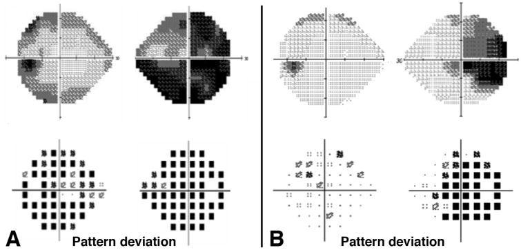
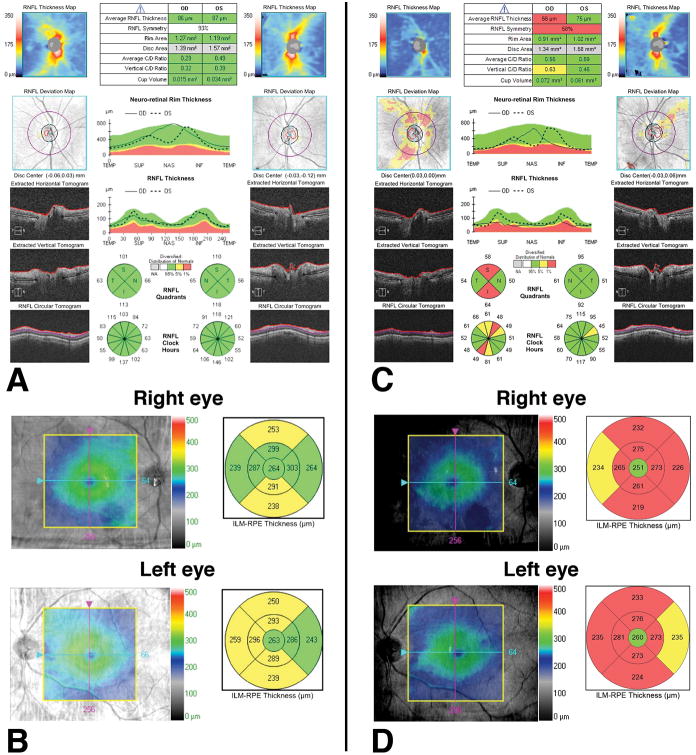
Background: We describe a 75-year-old woman who experienced vision loss in her left eye due to biopsy-proven giant cell arteritis (GCA). She subsequently developed pachymeningitis causing refractory headaches and bilateral optic neuropathy and maculopathy.
Methods: Case report with literature review.
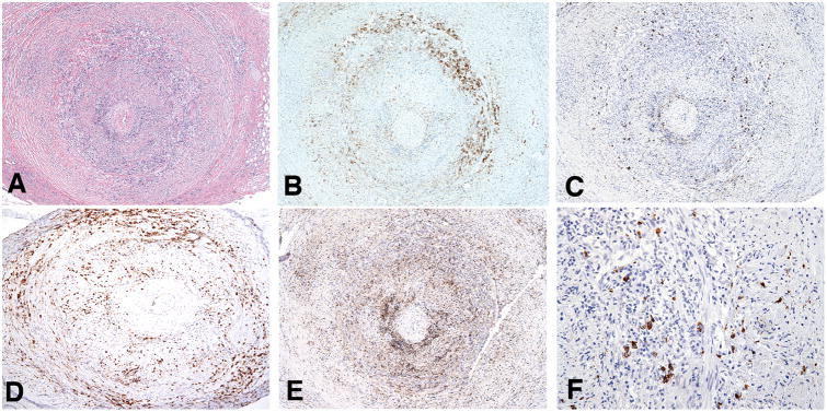
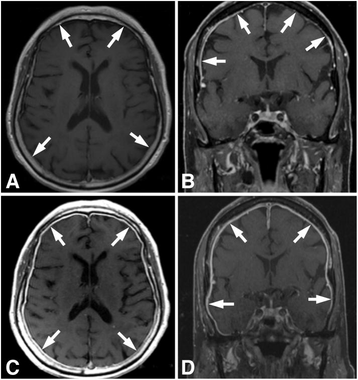
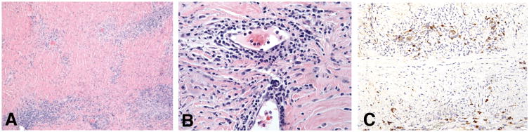
Results: Eighteen months after the initial diagnosis of GCA, imaging studies in our patient demonstrated pachymeningeal enhancement, and meningeal biopsy confirmed lymphoplasmacytic tissue infiltrates with low frequencies of IgG4+ plasma cells. Laboratory investigation revealed the presence of 3 antiretinal antibodies and antimyeloperoxidase antibodies, consistent with autoimmune retinopathy. Treatment with B-cell-depleting anti-CD20 antibodies suppressed meningeal inflammation and prevented further vision loss.
Conclusions: This case illustrates that bilateral vision loss and chronic headaches in patients with GCA may result from retina-directed autoimmunity and pachymeningitis.
Conflict of interest statement
Conflict of interest statement: The authors have no conflicts of interest to disclose.
Figures
Similar articles
-
Temporal arteritis with focal pachymeningitis: a deceptive association.Nagoya J Med Sci. 2020 Feb;82(1):143-150. doi: 10.18999/nagjms.82.1.143. Nagoya J Med Sci. 2020. PMID: 32273643 Free PMC article. Review.
-
Optic Neuropathy with Headache and Palpable Temporal Arteries Due to Hypertrophic Pachymeningitis Rather than Giant Cell Arteritis.Ocul Immunol Inflamm. 2022 Aug;30(6):1515-1518. doi: 10.1080/09273948.2021.1881561. Epub 2021 Apr 1. Ocul Immunol Inflamm. 2022. PMID: 33793376
-
Vision loss in giant cell arteritis: case-based review.Rheumatol Int. 2022 Oct;42(10):1855-1862. doi: 10.1007/s00296-022-05160-x. Epub 2022 Jun 21. Rheumatol Int. 2022. PMID: 35727336 Review.
-
Acute monocular visual loss in carcinomatous hypertrophic pachymeningitis mimicking giant cell arteritis.Rheumatol Int. 2006 May;26(7):683-4. doi: 10.1007/s00296-005-0049-4. Epub 2005 Dec 9. Rheumatol Int. 2006. PMID: 16341701
-
Unilateral proptosis resulting from giant-cell arteritis.J Am Optom Assoc. 1999 Jul;70(7):443-9. J Am Optom Assoc. 1999. PMID: 10485174
Cited by
-
Temporal arteritis with focal pachymeningitis: a deceptive association.Nagoya J Med Sci. 2020 Feb;82(1):143-150. doi: 10.18999/nagjms.82.1.143. Nagoya J Med Sci. 2020. PMID: 32273643 Free PMC article. Review.
-
Neurologic manifestations of giant cell arteritis.J Neurol. 2022 Jul;269(7):3430-3442. doi: 10.1007/s00415-022-10991-6. Epub 2022 Feb 6. J Neurol. 2022. PMID: 35124749 Review.
-
Role of temporal artery resection in Horton's arteritis (Review).Exp Ther Med. 2021 Oct;22(4):1099. doi: 10.3892/etm.2021.10533. Epub 2021 Aug 2. Exp Ther Med. 2021. PMID: 34504553 Free PMC article. Review.
References
Publication types
MeSH terms
Substances
Grants and funding
LinkOut - more resources
Full Text Sources
Other Literature Sources
Medical
